

9000SMZ Vibratome
High-Precision Vibrating Microtome - The New Standard in Precision Tissue Slicing
- Overview
- Specifications
- Accessories
- Citations
- Related Products
Overview
There are 4 images available to view - click to enlarge and scroll through the product gallery.
- Over 40 years experience with >4000 vibrating microtome publications.
- 5-Year Warranty.
- Proven high-quality slices with high cell viability.
- No chatter marks.
- Variable blade frequency & amplitude to cater for all tissue types.
- Precise control of advance speed, adjustable during slicing, for the perfect slice.
- Measure & correct blade Z-axis deflection (down to 0.1µm), essential for quality slices.
- Slice window mode for fast, automatic sectioning.
- Clear visibility of tissue; positional adjustments can be made during slicing.
- Store up to 8 user slice profiles for easy recall.
- Slice Quality Control Validation Data.
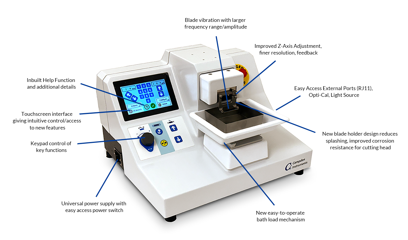
Advanced Vibratome Technology for Life Science and Biomedical Research
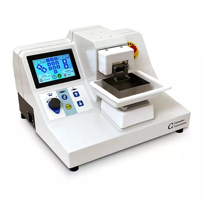
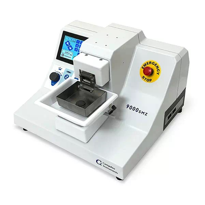
The Campden 9000SMZ Vibratome is our most advanced vibrating microtome, engineered for precise sectioning of live and fixed brain tissue. Trusted by neuroscience, cardiac, lung research, liver research, organoid research and plant research labs worldwide.
When your research depends on high-integrity tissue slices, compromise is not an option. The 9000SMZ Vibrating Microtome by Campden Instruments delivers unmatched control, slice quality, and tissue viability — making it the ideal vibratome for applications across electrophysiology, imaging, pathology, toxicology, and organ research.
Why Choose the 9000SMZ Vibrating Microtome?
Whether you're preparing slices of brain, kidney, lung, liver, retina, cardiac, pancreatic, brain organoids, tumour or plant tissue, the 9000SMZ provides the precision and reproducibility that today’s life sciences demand.
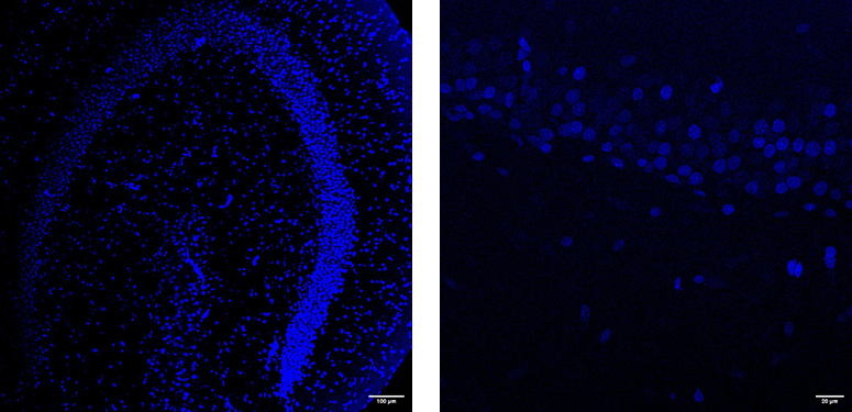
Mouse hippocampal slice: DAPI x10 + DAPI x40
One Vibratome, Endless Applications
The 9000SMZ is trusted in research involving:
- Neuroscience & electrophysiology
- Cancer and tumour microenvironment studies
- Renal, Precision Cut Liver Slices, Precision Cut Lung Slices, and cardiovascular research (living myocardial slices).
- Plant root and leaf sectioning
- Retinal and ocular tissue preparation
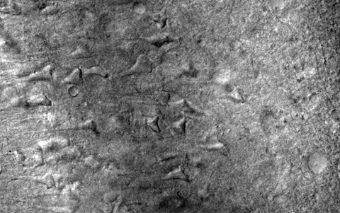
Entorhinal Cortex Slice from 9 month-old Rat
Superior Tissue Integrity Across Applications
- Ultra-low blade Z-axis deflection minimizes mechanical damage.
- Maintains structural and cellular viability in a wide range of soft tissues.
- Ideal for acute slicing, live tissue studies, ex vivo analysis, and long-term cultures
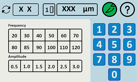
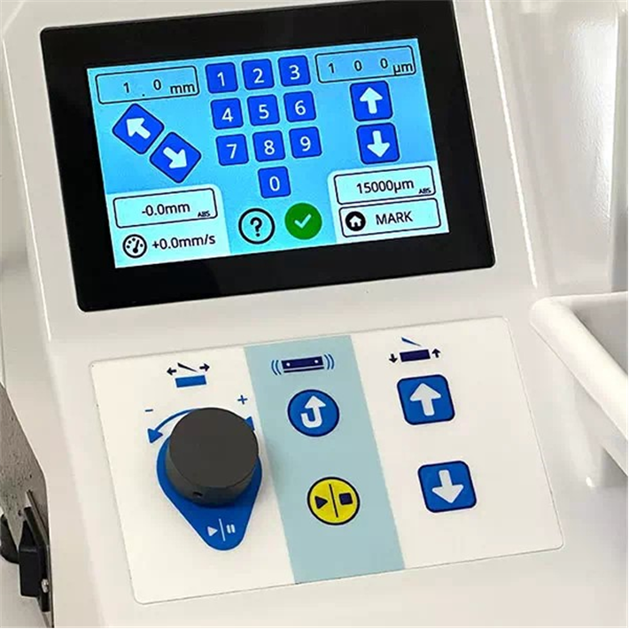
Parameters Screen
Smart, Automated Control System
- Touchscreen interface with programmable slicing profiles.
- Real-time feedback on blade amplitude, frequency, and advance rate.
- Minimizes training time and user-to-user variability.
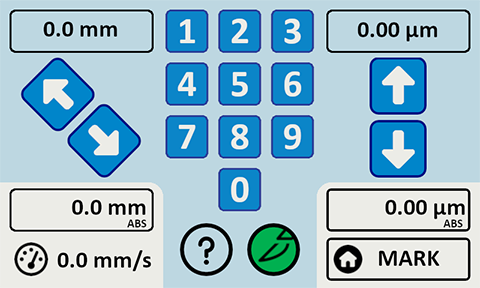
Move Screen
High-Precision Engineering
- Advance speed resolution of 10µm/s for reproducible section thickness
- Vibrating Blade holder has less than 0.1µm (100nm) deviation from path
- Compatible with a range of tissue sizes and embedding media
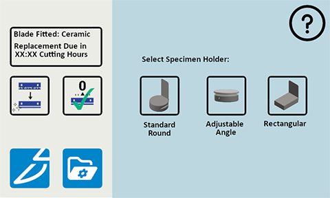
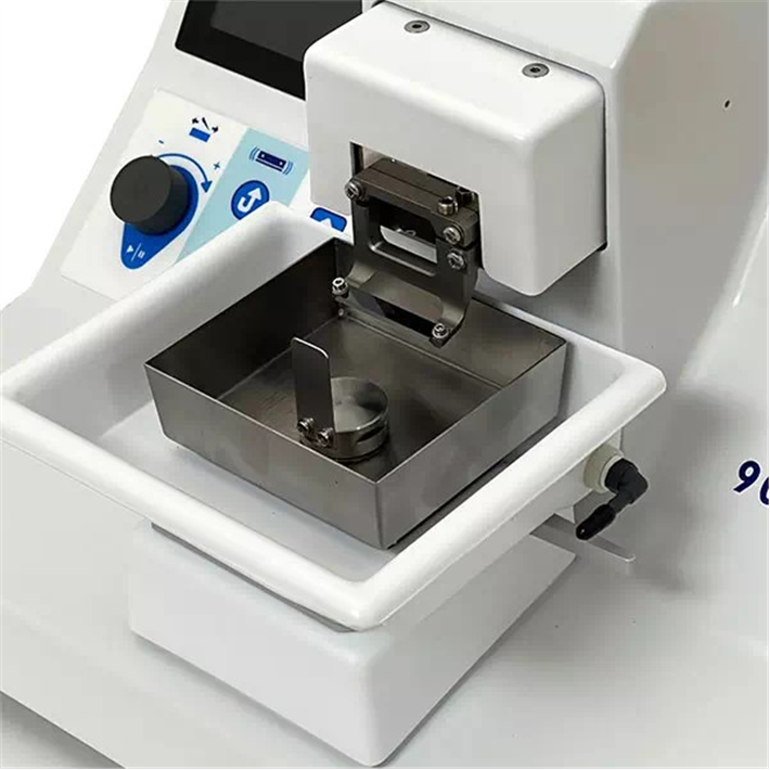
Preparation Screen
Built-In Tissue Protection
- Integrated cooling tray maintains physiological conditions
- Optional integrated Peltier cooling device
- Optimised for extended slicing sessions without compromising slice quality
Designed for Scientists, Backed by Innovation
Campden Instruments has collaborated with leading researchers across disciplines to build a vibratome that meets real-world lab challenges. With the 9000smz, you can expect:
- Cleaner slices
- Higher reproducibility
- Confidence from the very first cut
Customer Spotlights
Dr. Gareth Morris (University of Manchester and University College London) talks about his research and how the use of Brain Slices provides an invaluable, in vitro research tool.
Dr Kate Connor from Trinity College Dublin talks about her research into the development of clinically relevant cancer models and the use of brain slices for model validation.
Evolution: Meet the 9000smz Vibrating Microtome
Science
Oscillations in slices from 3 week-old mice*
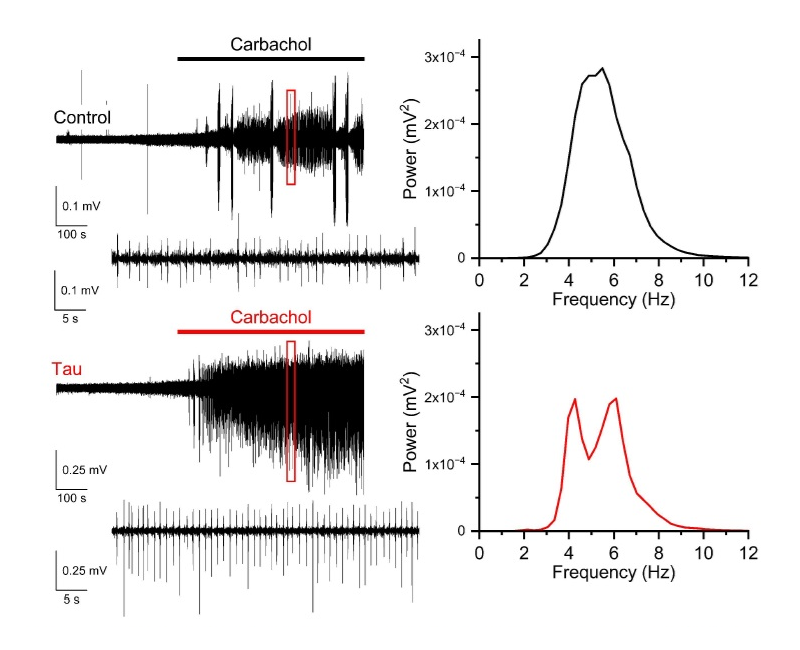
Neuronal oscillations in the theta frequency range (4 - 8 Hz) induced by the acetylcholine muscarinic receptor agonist carbachol (50 μM) in hippocampal area CA3 from 3 week-old C57BL/6J mice. Slices were either incubated in control aCSF or in aCSF supplemented with sonicated recombinant human tau preformed fibrils (133 nM; rPeptide; CF-1001-1). Left panels: raw electrophysiological traces showing induction of oscillations after carbachol, and 25 s of activity (red boxed region; lower traces). Right panels: spectrograms of theta activity induced by carbachol in both conditions.
Oscillations in slices from 9 month-old rats
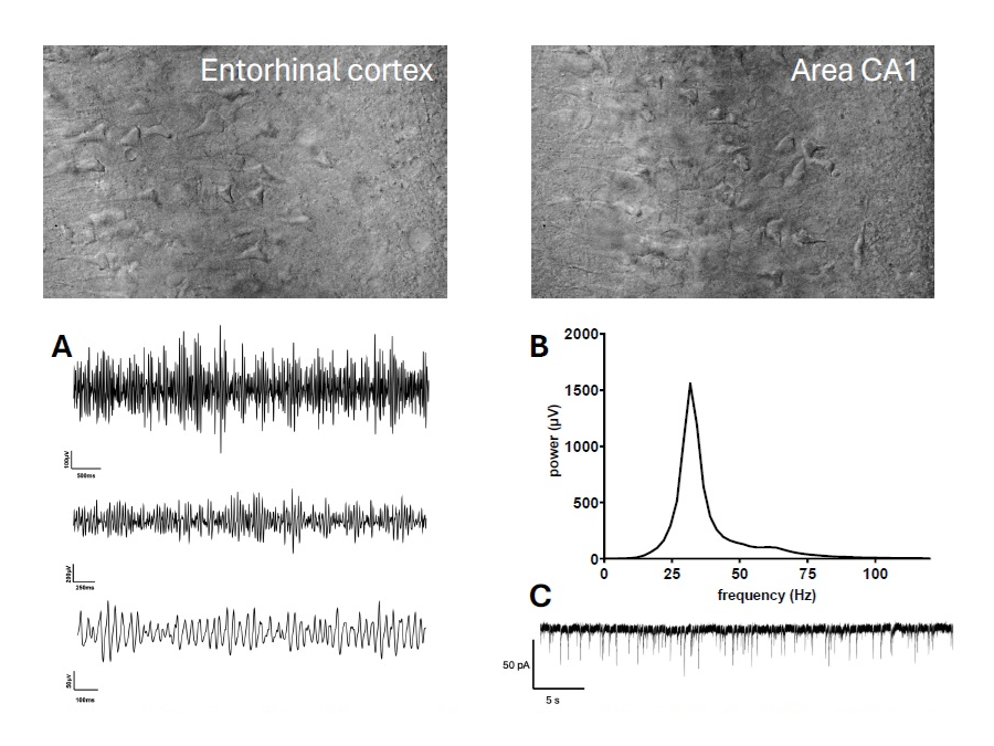
Improved microtome design allows neuronal preservation and physiological oscillations in older rats (9 months). Above: DIC images from entorhinal cortex and area CA1.
- Raw electrophysiological data at increasing magnification demonstrating characteristic spontaneous sawtooth gamma oscillations in LFP recordings (450 μm slice).
- Panel shows example FFT from LFP recording in hippocampus with no added drugs, with high power in gamma range and excellent coherence.
- Whole-cell patch-clamp recording from hippocampal cell with regular EPSCs and stable baseline (350 μm slice).
Immunohistochemistry*
Brainstem
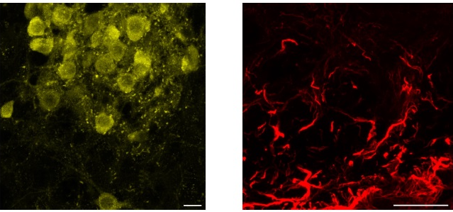
Maximum intensity projections of confocal images showing choline acetyltransferase (ChAT)+ neurons (left, yellow) and glial fibrillary acidic protein (GFAP)+ astroglia (right, red) in40 μm thick mouse brainstem sections. Scale bars = 20 μm
Hippocampus*
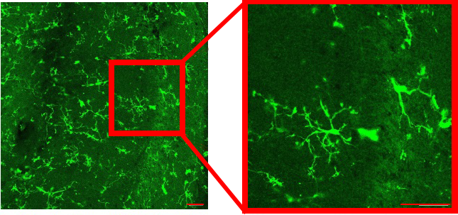
Left: Iba1 staining of LPS-stimulated microglia in the CA1 region of a 200 μm hippocampal slice. Right: highlighted region of interest. Scale bars = 50 μm
* Adwoa Boaten, Ben Hopkins, Tegan Lawrence, Beth Rees, Amol Bhandare, Stuart Greenhill, Gavin Woodhall, Mark Wall, Bruno Frenguelli School of Life Sciences, University of Warwick, Coventry, UK CV4 7AL. Institute of Health and Neurodevelopment, Aston University, Birmingham, UK B4 7ET


Request
Catalogue
Chat
Print