

BT-50MM-01
BiOTESTER: A fully equipped biaxial test system built specifically for biomaterials
- Overview
- Specifications
- Accessories
- Citations
- Related Products
Overview


There are 6 images available to view - click to enlarge and scroll through the product gallery.
Biotester Datasheet
/ Download as PDF
Uniaxial U-Stretch
/ Download as PDF
Why measure mechanical properties?
/ Download as PDF
Stress-strain in human sclera
/ Download as PDF
The BioTester provides researchers with an easy-to-use, affordable test instrument to characterize soft tissues and biomaterials. This biaxial test system captures and graphically displays live time, force, and synchronized video images for results analysis and verification. Data is easily exported to standard spreadsheet programs.
- High performance actuators (2 per axis) capable of μm positional resolution for accurate test motion.
- Inline overload-protected load cell on each axis
- High resolution CCD camera to collect time synchronized images for post test analysis
- Temperature controlled media bath
- Patented attachment system facilitates rapid and accurate specimen attachment
- Optional use of hook-and-suture or grip based attachment systems
- User-controlled test routines for multi-modal cyclic, simple, and relaxation testing over a wide range of strain rates
- Data output as a comma separated value text files for easy import into a variety of spread sheet and data analysis programs
- Simple USB connection to a Windows-based host computer
- Specimen Size: 3mm to 15mm
- Load Cell Capacities (N): 0.5, 1.5, 2.5, 5, 10, 23
- Load Cell Accuracies: 0.2% of capacity Max Displacement
- Rate: 10mm/s Image Rate: 15Hz
- Image Resolution: 2048 X 2048 pixels with CMOS camera
- Max. Temperature: 45°C
Sample Mounting Systems
The BioRake sample mounting system is CellScale’s patented method for attaching soft tissues and biomaterials.
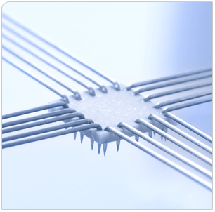
Each tine is electrochemically sharpened to easily pierce both the toughest and most delicate tissue samples. Each set is permanently attached to a common base to allow simultaneous puncture of all 20 attachment points. The BioRakes are magnetically mounted for easy removal for cleaning or replacement and for simple transition between BioRake, Balanced Pulley, and Clamp mounting systems.
To perform testing, samples are positioned and raised into place using the manual lift mechanism and pressure is applied to insert the hooks in the tissue. The sample is thus mounted and ready for analysis within a few seconds. The mounting is consistent, accurate and easy.
BioRakes are available with tine spacing ranging from 0.7mm to 2.2mm to accommodate specimens from 3 to 15mm in size.
The balanced pulley sample mounting system is CellScale’s attachment method for ensuring zero shear stress during biaxial testing.
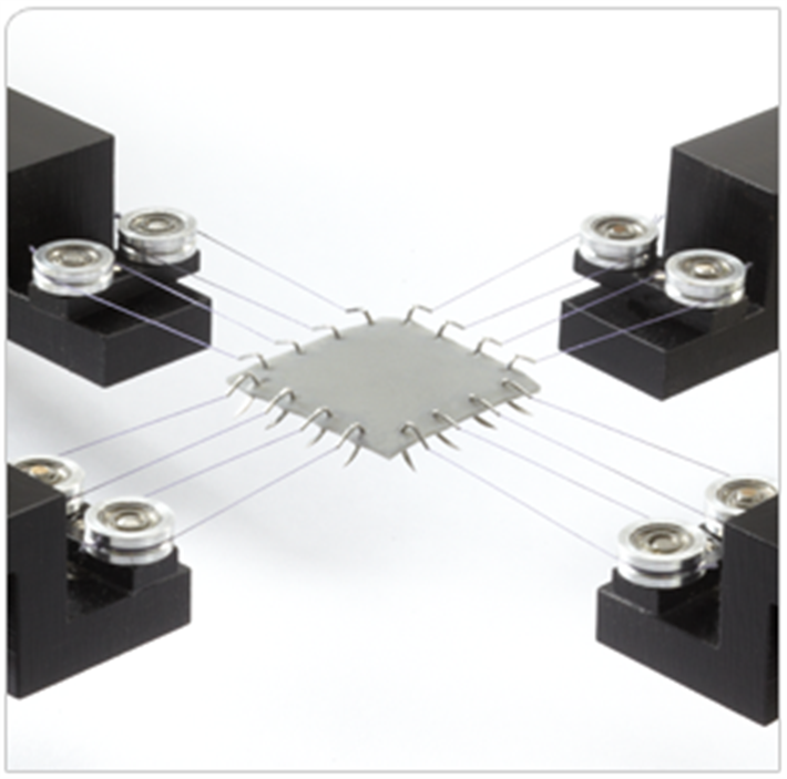
Two double-ended custom suture hooks are used to create 4 attachment points on each side of the specimen. A two-stage stainless steel pulley mechanism ensures that each of the sutures is held at the same tension during the test.
The pulley mechanisms are magnetically mounted for easy removal for cleaning and for simple transition between BioRake, Balanced Pulley, and Clamp mounting systems.
The clamp sample mounting system is CellScale’s attachment method for testing to failure.
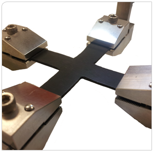
Using a cruciform specimen allows the attachment sites, which are inherently weaker than the base material, to be moved away from the gauge area of the specimen. The clamps allow the specimen to be loaded easily and held securely.
The stainless steel clamping mechanisms are mounted over the same brackets used for the other attachment systems to allow for fast and easy transition between BioRake, Balanced Pulley, and Clamp mounting systems.
Custom clamp designs can also be made to tailor the clamp force and clamping surface to your tissue.
Video Overviews
Specifications
| Force Capacity | 500, 100, 2500, 5000mN, 10N, 23N |
| Force Accuracy | 0.2% of force capacity |
| Max. Elongation Rate | 10mm/s |
| Max. Strain Rate (5mm specimen) | 200%/s |
| Spatial Resolution (Actuator) | >0.1μm |
| Spatial Accuracy (Acuator) | 10μm |
| Spatial Resolution (Image Analysis) | 1/8 pixel |
| Max. Force Data Rate | 100Hz |
| Image Rate | 1280 x 960 -15Hz |
Accessories
Citations
Caggiano, Laura & Holmes, Jeffrey. (2021). A Comparison of Fiber Based Material Laws for Myocardial Scar. Journal of Elasticity. 145. 1-17. 10.1007/s10659-021-09845-5.
Laurence, Devin & Homburg, Hannah & Tang, Qinggong & Fung, Kar-Ming & Bohnstedt, Bradley & Holzapfel, Gerhard & Lee, Chung-Hao. (2021). A pilot study on biaxial mechanical, collagen microstructural, and morphological characterizations of a resected human intracranial aneurysm tissue. Scientific Reports. 11. 10.1038/s41598-021-82991-x.
Leonov, D. & Spirina, Yu & Yatsenko, A. & Kushnarev, Vladimir & Ustinov, E. & Barannikov, S. (2021). Advanced 3D Bioprinting Technologies. Cell and Tissue Biology. 15. 616-627. 10.1134/S1990519X21060134.
Borem, Ryan & Madeline, Allison & Theos, Chris & Vela, Ricardo & Garon, Alex & Gill, Sanjitpal & Mercuri, Jeremy. (2021). Angle-ply scaffold supports annulus fibrosus matrix expression and remodeling by mesenchymal stromal and annulus fibrosus cells. Journal of Biomedical Materials Research Part B: Applied Biomaterials. 10.1002/jbm.b.34980.
McClarty, Davis & Ouzounian, Maral & Tang, Mingyi & Eliathamby, Daniella & Romero, David & Nguyen, Elsie & Simmons, Craig & Amon, Cristina & Chung, Jennifer. (2021). Ascending aortic aneurysm haemodynamics are associated with aortic wall biomechanical properties. European Journal of Cardio-Thoracic Surgery. 10.1093/ejcts/ezab471.
Ndlovu, Zwelihle & Desai, Dawood & Pandelani, Thanyani & Ngwangwa, Harry & Nemavhola, Fulufhelo. (2021). Biaxial estimation of biomechanical constitutive parameters of passive porcine sclera soft tissue. 10.31224/osf.io/h27wp.
Filippo Valente, Matt S. Hepburn, Jingyu Chen, Ana A. Aldana, Benjamin J. Allardyce, Sajjad Shafei, Barry J. Doyle, Brendan F. Kennedy, Rodney J. Dilley, (2021). Bioprinting silk fibroin using two-photon lithography enables control over the physico-chemical material properties and cellular response. Bioprinting. 25. e00183. 10.1016/j.bprint.2021.e00183.
Lomboni, David & Steeves, Alexander & Schock, Sarah & Bonetti, Lorenzo & De Nardo, Luigi & Variola, Fabio. (2021). Compounded topographical and physicochemical cueing by micro-engineered chitosan substrates on rat dorsal root ganglion neurons and human mesenchymal stem cells. Soft Matter. 17. 10.1039/D0SM02170A.
Xu, Liangpeng & Yang, Fan & Ge, Yao & Guo, Gaoyang & Wang, Yunbing. (2021). Crosslinking porcine aortic valve by radical polymerization for the preparation of BHVs with improved cytocompatibility, mild immune response, and reduced calcification. Journal of Biomaterials Applications. 35. 088532822098406. 10.1177/0885328220984066.
Tang, Mingyi & Eliathamby, Daniella & Ouzounian, Maral & Simmons, Craig & Chung, Jennifer (2021). Dependency of energy loss on strain rate, strain magnitude and preload: Towards development of a novel biomarker for aortic aneurysm dissection risk. Journal of the Mechanical Behavior of Biomedical Materials. 124. 104736. 10.1016/j.jmbbm.2021.104736.
Laurence, Devin & Lee, Chung-Hao. (2021). Determination of a Strain Energy Density Function for the Tricuspid Valve Leaflets Using Constant Invariant-Based Mechanical Characterizations. Journal of Biomechanical Engineering. 143. 10.1115/1.4052612.
Ngwangwa, Harry & Nemavhola, Fulufhelo & Pandelani, Thanyani & Msibi, Makhosasana & Mabuda, Israel & Davies, Neil & Franz, Thomas. (2021). Determination of Cross-directional and Cross-Wall Variations of Passive Biaxial Mechanical Properties of Rat Myocardium. 10.20944/preprints202109.0244.v1.
Aldana, Agustina & Valente, Filippo & Dilley, Rodney & Doyle, Barry (2021). Development of 3D bioprinted GelMA-alginate hydrogels with tunable mechanical properties. Bioprinting. 21. e00105. 10.1016/j.bprint.2020.e00105.
Hu, Cheng & Long, Linyu & Cao, Juan & Zhang, Shumang & Wang, Yunbing. (2021). Dual-crosslinked mussel-inspired smart hydrogels with enhanced antibacterial and angiogenic properties for chronic infected diabetic wound treatment via pH-responsive quick cargo release. Chemical Engineering Journal. 411. 128564. 10.1016/j.cej.2021.128564.
Ross, Colton & Laurence, Devin & Echols, Allyson & Babu, Anju & Gu, Tingting & Duginski, Grace & Johns, Cortland & Mullins, Brennan & Casey, Katherine & Laurence, Keely & Zhao, Yan & Amini, Rouzbeh & Fung, Kar-Ming & Mir, Arshid & Burkhart, Harold & Wu, Yi & Holzapfel, Gerhard & Lee, Chung-Hao. (2021). Effects of enzyme-based removal of collagen and elastin constituents on the biaxial mechanical responses of porcine atrioventricular heart valve anterior leaflets. Acta Biomaterialia. 135. 10.1016/j.actbio.2021.08.043.
Ross, Colton & Mullins, Brennan & Hillshafer, Clare & Mir, Arshid & Burkhart, Harold & Lee, Chung-Hao. (2021). Evaluation of affine fiber kinematics in porcine tricuspid valve leaflets using polarized spatial frequency domain imaging and planar biaxial testing. Journal of Biomechanics. 123. 110475. 10.1016/j.jbiomech.2021.110475.
Pillalamarri, Narasimha & Patnaik, Sourav & Piskin, Senol & Gueldner, Pete & Finol, Ender. (2021). Ex Vivo Regional Mechanical Characterization of Porcine Pulmonary Arteries. Experimental Mechanics. 61. 1-19. 10.1007/s11340-020-00678-2.
Nemavhola, Fulufhelo & Ngwangwa, Harry & Pandelani, Thanyani. (2021). Experimental analysis and biaxial biomechanical behaviour of ex-vivo sheep trachea. 10.1101/2021.11.26.470180.
Durbak, Emily & Tarraf, Samar & Gillespie, Callan & Germano, Emídio & Cikach, Frank & Blackstone, Eugene & Emerton, Kelly & Colbrunn, Robb & Bellini, Chiara & Roselli, Eric. (2021). Ex-vivo Biaxial Load Testing Analysis of Aortic Biomechanics Demonstrates Variation in Elastic Energy Distribution Across the Aortic Zone Zero. The Journal of Thoracic and Cardiovascular Surgery. 10.1016/j.jtcvs.2021.09.071.
Nemavhola, Fulufhelo & Pandelani, Thanyani & Ngwangwa, Harry. (2021). Fitting Of Hyperelastic Constitutive Models In Different Sheep Heart Regions Based On Biaxial Mechanical Properties. 10.1101/2021.10.28.466240.
Ramburrun, Poornima & Kumar, Pradeep & Ndobe, Elias & Choonara, Yahya. (2021). Gellan-Xanthan Hydrogel Conduits with Intraluminal Electrospun Nanofibers as Physical, Chemical and Therapeutic Cues for Peripheral Nerve Repair. International Journal of Molecular Sciences. 22. 11555. 10.3390/ijms222111555.
Hudson, Luke & Laurence, Devin & Lau, Hunter & Mullins, Brennan & Doan, Deenna & Lee, Chung-Hao. (2021). Linking collagen fiber architecture to tissue-level biaxial mechanical behaviors of porcine semilunar heart valve cusps. Journal of the Mechanical Behavior of Biomedical Materials. 125. 104907. 10.1016/j.jmbbm.2021.104907.
Surman, Tim & O'Rourke, Dermot & Reynolds, Karen & Edwards, J. & Worthington, M. (2021). M06 The Unique Tissue Biomechanics of the Thoracic Aorta. What are the Greatest Areas of Weakness and Where Should we Focus Repair?. Heart, Lung and Circulation. 30. S4. 10.1016/j.hlc.2021.03.015.
Singh, B. & Singh, G. & Chard, R. & Nicholson, I. (2021). M08 Perioperative and Midterm Outcomes of Aortic Root Enlargements Compared to Conventional and Rapid Deployment Prosthesis. Heart, Lung and Circulation. 30. S4-S5. 10.1016/j.hlc.2021.03.017.
Walsh, Darragh & Ross, Aisling & Newport, David & Zhou, Zhou & Kearns, Jamie & Fearon, Conor & Lorigan, Jennifer & Mulvihill, John. (2021). Mechanical Characterisation of the Human Dura Mater, Falx Cerebri and Superior Sagittal Sinus. Acta Biomaterialia. 134. 10.1016/j.actbio.2021.07.043.
Cunnane, Eoghan & Davis, NIALL & Cunnane, Connor & Lorentz, Katherine & Ryan, Alan & Hess, Jochen & Weinbaum, Justin & Walsh, Michael & O’Brien, Fergal & Vorp, David. (2021). Mechanical, compositional and morphological characterisation of the human male urethra for the development of a biomimetic tissue engineered urethral scaffold. Biomaterials. 269. 120651. 10.1016/j.biomaterials.2021.120651.
Maleckis, Kaspars & Kamenskiy, Alexey & Lichter, Eliezer & Oberley-Deegan, Rebecca & Dzenis, Yuris & Mactaggart, Jason. (2021). Mechanically Tuned Vascular Graft Demonstrates Rapid Endothelialization and Integration Into the Porcine Iliac Artery Wall. Acta Biomaterialia. 125. 10.1016/j.actbio.2021.01.047.
Morningstar, Jordan & Gensemer, Cortney & Moore, Reece & Fulmer, Diana & Beck, Tyler & Wang, Christina & Moore, Kelsey & Guo, Lilong & Sieg, Franz & Nagata, Yasufumi & Bertrand, Philippe & Spampinato, Ricardo & Glover, Janiece & Poelzing, Stephen & Gourdie, Robert & Watts, Kelsey & Richardson, William & Levine, Robert & Borger, Michael & Norris, Russell. (2021). Mitral Valve Prolapse Induces Regionalized Myocardial Fibrosis. Journal of the American Heart Association. 10. 10.1161/JAHA.121.022332.
Ahmad, Dilshad & Ajaj, Rafic. (2021). Multiaxial Mechanical Characterization of Latex Skin for Morphing Wing Application. Polymer Testing. 10.1016/j.polymertesting.2021.107408.
Zheng, Cheng & Ding, Kailei & Huang, Xueyu & Li, Meiling & Wu, Bingang & Lei, Yang & Wang, Yunbing. (2021). Nonglutaraldehyde crosslinked bioprosthetic heart valves based on 2-isocyanatoethyl methacrylate crosslinked porcine pericardium with improved properties of stability, cytocompatibility and anti-calcification. Composites Part B: Engineering. 230. 109504. 10.1016/j.compositesb.2021.109504.
Nemavhola, Fulufhelo & Ngwangwa, Harry & Davies, Neil & Franz, Thoams. (2021). Passive Biaxial Tensile Dataset of Three Main Rat Heart Myocardia: Left Ventricle, Mid-Wall and Right Ventricle. 10.20944/preprints202108.0153.v1.
Liu, Hailong & Jain, Shubham & Ahlinder, Astrid & Fuoco, Tiziana & Gasser, Thomas & Finne-Wistrand, Anna. (2021). Pliable, Scalable, and Degradable Scaffolds with Varying Spatial Stiffness and Tunable Compressive Modulus Produced by Adopting a Modular Design Strategy at the Macrolevel. ACS Polymers Au. XXXX. 10.1021/acspolymersau.1c00013.
Ross, Colton & Hsu, Ming-Chen & Baumwart, Ryan & Mir, Arshid & Burkhart, Harold & Holzapfel, Gerhard & Wu, Yi & Lee, Chung-Hao. (2021). Quantification of load-dependent changes in the collagen fiber architecture for the strut chordae tendineae-leaflet insertion of porcine atrioventricular heart valves. Biomechanics and Modeling in Mechanobiology. 20. 10.1007/s10237-020-01379-4.
Ndlovu, Zwelihle & Desia, Dawood & Nemavhola, Fulufhelo & Ngwangwa, Harry. (2021). Sheep Sclera Soft Tissue Subjected to Mechanical Equi-Biaxial Testing. 10.20944/preprints202108.0388.v1.
Quince, Zachery & Alonso-Caneiro, David & Read, Scott & Collins, Michael. (2021). Static compression optical coherence elastography to measure soft contact lens mechanical properties. Biomedical Optics Express. 12. 10.1364/BOE.419344.
Nemavhola, Fulufhelo. (2021). Study of biaxial mechanical properties of the passive pig heart: material characterisation and categorisation of regional differences. International Journal of Mechanical and Materials Engineering. 16. 10.1186/s40712-021-00128-4.
Hu, Yingbing & Huang, Yu & Chen, Yun & Ye, Cheng & Wei, Wei & Feng, Yun & Mi, Shengli. (2021). Study on patterned photodynamic cross-linking for keratoconus. Experimental Eye Research. 204. 108450. 10.1016/j.exer.2021.108450.
Cai, Li & Zhang, Ruihang & Li, Yiqiang & Guangyu, Zhu & Ma, Xingshuang & Wang, Yongheng & Luo, Xiaoyu & Gao, Hao. (2021). The Comparison of Different Constitutive Laws and Fiber Architectures for the Aortic Valve on Fluid–Structure Interaction Simulation. Frontiers in Physiology. 12. 682893. 10.3389/fphys.2021.682893.
Meador, William & Zhou, Jennifer & Malinowski, Marcin & Jazwiec, Tomasz & Calve, Sarah & Timek, Tomasz & Rausch, Manuel. (2021). The effects of a simple optical clearing protocol on the mechanics of collagenous soft tissue. Journal of Biomechanics. 122. 110413. 10.1016/j.jbiomech.2021.110413.
Barrett, Jeff & Fewster, Kayla & Cudlip, Alan & Dickerson, Clark & Callaghan, Jack. (2021). The rate of tendon failure in a collagen fibre recruitment-based model. Journal of the Mechanical Behavior of Biomedical Materials. 115. 104273. 10.1016/j.jmbbm.2020.104273.
Sawadkar, Prasad & Nandin, Mandakhbayar & Patel, Kapil & Buitrago, Jennifer & Kim, Tae & Rajasekar, Poojitha & Lali, Ferdinand & Kyriakidis, Christos & Rahmani, Benyamin & Mohanakrishnan, Jeviya & Dua, Rishbha & Greco, Karin & Lee, Jung-Hwan & Kim, Hae-Won & Knowles, Jonathan & García-Gareta, Elena. (2021). Three dimensional porous scaffolds derived from collagen, elastin and fibrin proteins orchestrate adipose tissue regeneration. Journal of Tissue Engineering. 12. 1-17. 10.1177/20417314211019238.
Nemavhola, Fulufhelo & Ngwangwa, Harry & Pandelani, Thanyani & Davies, Neil & Franz, Thomas. (2021). Understanding regional mechanics of rat myocardia by fitting hyperelatsic models. 10.21203/rs.3.rs-957393/v1.
Yang, Fan & Xu, Liangpeng & Guo, Gaoyang & Wang, Yunbing. (2021). Visible light–induced cross-linking of porcine pericardium for the improvement of endothelialization, anti-tearing, and anticalcification properties. Journal of Biomedical Materials Research Part A. 110. 10.1002/jbm.a.37263.
Lan, Xiaorong & Zhao, Qianting & Zhang, Jiayi & Lei, Yang & Wang, Yunbing. (2020). A combination of hydrogen bonding and chemical covalent crosslinking to fabricate a novel swim-bladder-derived dry heart valve material yields advantageous mechanical and biological properties. Biomedical Materials. 16. 10.1088/1748-605X/abb616.
A. Cudlip (2020). A combined in vivo and in vitro approach to assess supraspinatus activation and tissue responses to arm elevation demands
Sang, Cyril & Kallmes, D. & Kadirvel, R. & Durka, Micheal & Ding, Y-H & Dai, D. & Watkins, S. & Robertson, Anne (2020). Adaptive Remodeling in the Elastase-Induced Rabbit Aneurysms. Experimental Mechanics. 61. 10.1007/s11340-020-00671-9.
Sharifi Kia, Danial & Benza, Evan & Bachman, Timothy & Tushak, Claire & Kim, Kang & Simon, Marc. (2020). Angiotensin Receptor-Neprilysin Inhibition Attenuates Right Ventricular Remodeling in Pulmonary Hypertension. Journal of the American Heart Association. 9. 10.1161/JAHA.119.015708.
Schaefer, Douglas & Khan, Sehroon & Nadir, Sadia & Dong, Yang & Mortimer, Peter & Gui, Heng & Khan, Afsar & Yu, Mingming & Iqbal, Shahid & Sheng, Jun & Xu, Jianchu. (2020). Biodegradation of polyester polyurethane by Aspergillus flavus G10. 10.1101/2020.06.25.170654.
Chung, Jennifer & Wong, Edwin & Tang, Mingyi & Eliathamby, Daniella & Forbes, Thomas & Butany, Jagdish & Simmons, Craig & Ouzounian, Maral. (2020). Biomechanics of Aortic Dissection: A Comparison of Aortas Associated With Bicuspid and Tricuspid Aortic Valves. Journal of the American Heart Association. 9. 10.1161/JAHA.120.016715.
Ahn, Seungkuk & Chantre, Christophe & Ardona, Herdeline Ann & Gonzalez, Grant & Campbell, Patrick & Parker, Kevin. (2020). Biomimetic and estrogenic fibers promote tissue repair in mice and human skin via estrogen receptor β. Biomaterials. 255. 120149. 10.1016/j.biomaterials.2020.120149.
Virgilio, Kelley & Jones, Brian & Miller, Emily & Ghajar-Rahimi, Elnaz & Martin, Kyle & Peirce, Shayn & Blemker, Silvia. (2020). Computational Models Provide Insight into In Vivo Studies and Reveal the Complex Role of Fibrosis in mdx Muscle Regeneration. Annals of Biomedical Engineering. 49. 10.1007/s10439-020-02566-1.
Jadidi, Majid & Sherifova, Selda & Sommer, Gerhard & Kamenskiy, Alexey & Holzapfel, Gerhard. (2020). Constitutive modeling using structural information on collagen fiber direction and dispersion in human superficial femoral artery specimens of different ages. Acta Biomaterialia. 121. 10.1016/j.actbio.2020.11.046.
Wu, Binggang & Zheng, Cheng & Ding, Kailei & Huang, Xueyu & Li, Meiling & Zhang, Shumang & Lei, Yang & Guo, Yingqiang & Wang, Yunbing. (2020). Cross-Linking Porcine Pericardium by 3,4-Dihydroxybenzaldehyde: A Novel Method to Improve the Biocompatibility of Bioprosthetic Valve. Biomacromolecules. 22. 10.1021/acs.biomac.0c01554.
Miar, Solaleh & Dion, Gregory & Montelongo, Sergio & Ong, Joo & Bizios, Rena & Guda, Teja. (2020). Development of a Bioinspired, Self-Adhering, and Drug-Eluting Laryngotracheal Patch. The Laryngoscope. 131. 10.1002/lary.29182.
Desyatova, Anastasia & Mactaggart, Jason & Kamenskiy, Alexey. (2020). Effects of longitudinal pre-stretch on the mechanics of human aorta before and after thoracic endovascular aortic repair (TEVAR) in trauma patients. Biomechanics and Modeling in Mechanobiology. 19. 10.1007/s10237-019-01217-2.
Duijvelshoff, Renee & Di Luca, Andrea & Van Haaften, Eline & Dekker, Sylvia & Söntjens, Serge & Janssen, Henk & Smits, Anthal & Dankers, Patricia & Bouten, Carlijn. (2020). Inconsistency in Graft Outcome of Bilayered Bioresorbable Supramolecular Arterial Scaffolds in Rats. Tissue Engineering Part A. 27. 10.1089/ten.TEA.2020.0185.
Yang, Fan & He, Haiyang & Xu, Liangpeng & Jin, Linhe & Guo, Gaoyang & Wang, Yunbing. (2020). Inorganic-polymerization crosslinked tissue-siloxane hybrid as potential biomaterial for bioprosthetic heart valves. Journal of Biomedical Materials Research Part A. 109. 10.1002/jbm.a.37061.
Jett, Samuel & Hudson, Luke & Baumwart, Ryan & Bohnstedt, Bradley & Mir, Arshid & Burkhart, Harold & Holzapfel, Gerhard & Wu, Yi & Lee, Chung-Hao. (2020). Load-dependent collagen fiber architecture data of representative bovine tendon and mitral valve anterior leaflet tissues as quantified by an integrated opto-mechanical system. Data in Brief. 28. 105081. 10.1016/j.dib.2019.105081.
Yang, Li & Wang, Yunbing & Ding, Kailei & Xiao, Chaonan & Lei, Yang & Du, Mingchun & Pan, Dengke & Guo, Xiumei. (2020). Low Immunogenicity Pericardia from Gene Knockout Pigs as Biological Valve Materials. SSRN Electronic Journal. 10.2139/ssrn.3674057.
He, Qizhi & Laurence, Devin & Lee, Chung-Hao & Chen, Jiun-Shyan. (2020). Manifold learning based data-driven modeling for soft biological tissues. Journal of Biomechanics. 117. 110124. 10.1016/j.jbiomech.2020.110124.
Chen, Shengda & Sari, Candra & Gao, Hao & Lei, Yang & Segers, Patrick & Beule, Matthieu & Wang, Guixue & Ma, Xingshuang. (2020). Mechanical and morphometric study of mitral valve chordae tendineae and related papillary muscle. Journal of the Mechanical Behavior of Biomedical Materials. 111. 104011. 10.1016/j.jmbbm.2020.104011.
Walsh, Darragh & Lynch, James & Connor, David & Newport, David & Mulvihill, John. (2020). Mechanical and structural characterisation of the dural venous sinuses. Scientific Reports. 10. 10.1038/s41598-020-78694-4.
Whittal, Mitchel & Molladavoodi, Sara & Zwambag, Derek & Millecamps, Magali & Stone, Laura & Gregory, Diane. (2020). Mechanical Consequence of Induced Intervertebral Disc Degeneration in the SPARC-Null Mouse. Journal of Biomechanical Engineering. 143. 10.1115/1.4047995.
Ross, Colton & Laurence, Devin & Hsu, Ming-Chen & Baumwart, Ryan & Zhao, Daniel & Mir, Arshid & Burkhart, Harold & Holzapfel, Gerhard & Wu, Yi & Lee, Chung-Hao. (2020). Mechanics of Porcine Heart Valves’ Strut Chordae Tendineae Investigated as a Leaflet–Chordae–Papillary Muscle Entity. Annals of Biomedical Engineering. 48. 10.1007/s10439-020-02464-6.
Yang, Fan & Xu, Liangpeng & Kuang, Dajun & Ge, Yao & Guo, Gaoyang & Wang, Yunbing. (2020). Polyzwitterion-crosslinked hybrid tissue with antithrombogenicity, endothelialization, anticalcification properties. Chemical Engineering Journal. 410. 128244. 10.1016/j.cej.2020.128244.
Yang, Li & Huang, Xueyu & Deng, Lu & Ma, Xiaoxiao & Jiang, Honglin & Ning, Qinggong & Liang, Zhen & Lei, Yang & Wang, Yunbing. (2020). Pre-mounted dry TAVI valve with improved endothelialization potential using REDV-loaded PEGMA hydrogel hybrid pericardium. Journal of Materials Chemistry B. 8. 10.1039/C9TB00879A.
Aldosary, Ghada & Tse, Tabitha & Arnaout, Angel & Caudrelier, Jean-Michel & Czyrnyj, Catriona & Romain, Ron & Mclean, Linda & Foottit, Claire & Vandervoort, Eric & Belec, Jason. (2020). Radiological, dosimetric and mechanical properties of a deformable breast phantom for radiation therapy and surgical applications. Biomedical Physics & Engineering Express. 6. 10.1088/2057-1976/ab834a.
Barbour, Kaitlyn & Huang, Hsiao-Ying. (2020). Strain effects on collagen proteolysis in heart valve tissues. Mechanics of Time-Dependent Materials. 24. 10.1007/s11043-019-09410-7.
Yang, Li & Xie, Shuang & Ding, Kailei & Lei, Yang & Wang, Yunbing. (2020). The study of dry biological valve crosslinked with a combination of carbodiimide and polyphenol. Regenerative Biomaterials. 8. 10.1093/rb/rbaa049.
Meador, William & Mathur, Mrudang & Sugerman, Gabriella & Malinowski, Marcin & Jazwiec, Tomasz & Wang, Xinmei & Lacerda, Carla & Timek, Tomasz & Rausch, Manuel. (2020). The tricuspid valve also maladapts as shown in sheep with biventricular heart failure. eLife. 9. 10.7554/eLife.63855.
Meador, William & Mathur, Mrudang & Sugerman, Gabriella & Malinowski, Marcin & Jazwiec, Tomasz & Wang, Xinmei & Lacerda, Carla & Timek, Tomasz & Rausch, Manuel. (2020). The tricuspid valve also maladapts: A multiscale study in sheep with biventricular heart failure. 10.1101/2020.09.03.278515.
Jin, Linhe & He, Haiyang & Yang, Fan & Xu, Liangpeng & Guo, Gaoyang & Wang, Yunbing. (2020). Tough pNAGA hydrogel hybridized porcine pericardium for the pre-mounted TAVI valve with improved anti-tearing properties and hemocompatibility. Biomedical Materials. 15. 10.1088/1748-605X/aba239.
Smith, Kieran & Mathur, Mrudang & Meador, William & Phillips-Garcia, B. & Sugerman, G. & Menta, A. & Jazwiec, T. & Malinowski, Marcin & Timek, T. & Rausch, Manuel. (2020). Tricuspid Chordae Tendineae Mechanics: Insertion Site, Leaflet, and Size-Specific Analysis and Constitutive Modelling. Experimental Mechanics. 61. 10.1007/s11340-020-00594-5.
Tavakoli, Javad & Raston, Colin & Ma, Yong & Tang, Youhong. (2020). Vortex fluidic mediated one-step fabrication of polyvinyl alcohol hydrogel films with tunable surface morphologies and enhanced self-healing properties. Science China Materials. 63. 10.1007/s40843-020-1301-y.
Meador, William & Mathur, Mrudang & Sugerman, Gabriella & Jazwiec, Tomasz & Malinowski, Marcin & Bersi, Matthew & Timek, Tomasz & Rausch, Manuel. (2019). A Detailed Mechanical and Microstructural Analysis of Ovine Tricuspid Valve Leaflets. Acta Biomaterialia. 102. 10.1016/j.actbio.2019.11.039.
S. Jett, Z. Schuermann, A. Mir, H. Burkhart, C-H. Lee (2019). An Integrated Opto-Mechanical System for Quantification of Dynamic Microstructure and Mechanics of Heart Valve Tissues. https://samjett.com/files/Jett_SB3C_Abstract.pdf
Kramer, Katherine & Ross, Colton & Laurence, Devin & Babu, Anju & Wu, Yi & Towner, Rheal & Mir, Arshid & Burkhart, Harold & Holzapfel, Gerhard & Lee, Chung-Hao. (2019). An investigation of layer-specific tissue biomechanics of porcine atrioventricular valve anterior leaflets. Acta Biomaterialia. 96. 10.1016/j.actbio.2019.06.049.
Sari, C.R., Chen, S., Lei, Y., Gao, H., Wang, G. and Ma, X. (2019). An Investigation of Regional Variations in the Biaxial Mechanical Properties of Porcine Mitral Valve. https://www.cnki.com.cn/Article/CJFDTotal-YISX2019S1202.htm
Duginski, Grace & Ross, Colton & Laurence, Devin & Johns, Cortland & Lee, Chung-Hao. (2019). An investigation of the effect of freezing storage on the biaxial mechanical properties of excised porcine tricuspid valve anterior leaflets. Journal of the Mechanical Behavior of Biomedical Materials. 101. 103438. 10.1016/j.jmbbm.2019.103438.
Ross, Colton & Laurence, Devin & Richardson, Jacob & Babu, Anju & Evans, Lauren & Beyer, Ean & Childers, Rachel & Wu, Yi & Towner, Rheal & Fung, Kar-Ming & Mir, Arshid & Burkhart, Harold & Holzapfel, Gerhard & Lee, Chung-Hao. (2019). An investigation of the glycosaminoglycan contribution to biaxial mechanical behaviours of porcine atrioventricular heart valve leaflets. Journal of The Royal Society Interface. 16. 20190069. 10.1098/rsif.2019.0069.
Ross, Colton & Laurence, Devin & Wu, Yi & Lee, Chung-Hao. (2019). Biaxial Mechanical Characterizations of Atrioventricular Heart Valves. Journal of Visualized Experiments. 10.3791/59170.
Patnaik, Sourav & Piskin, Senol & Pillalamarri, Narasimha & Romero, Gabriela & Escobar, Gladys & Sprague, Eugene & Finol, Ender. (2019). Biomechanical Restoration Potential of Pentagalloyl Glucose after Arterial Extracellular Matrix Degeneration. Bioengineering. 6. 58. 10.3390/bioengineering6030058.
Van Haaften, Eline & Wissing, T.B. & Kurniawan, Nicholas Agung & Smits, Anthal & Bouten, Carlijn. (2019). Human in vitro model of material-driven vascular regeneration reveals how cyclic stretch and shear stress differentially modulate inflammation and tissue formation. 10.1101/755157.
Van Haaften, Eline & Turnhout, Mark & Kurniawan, Nicholas Agung. (2019). Image-based analysis of uniaxial ring test for mechanical characterization of soft materials and biological tissues. Soft Matter. 15. 3353-3361. 10.1039/c8sm02343c.
Samuel V. Jett, Luke T. Hudson, Ryan Baumwart, Bradley N. Bohnstedt, Arshid Mir, Harold M. Burkhart, Gerhard A. Holzapfel, Yi Wu, Chung-Hao Lee (2019). Integration of polarized spatial frequency domain imaging (pSFDI) with a biaxial mechanical testing system for quantification of load-dependent collagen architecture in soft collagenous tissues. https://doi.org/10.1016/j.actbio.2019.11.028
Wissing, T.B. & Bonito, Valentina & Van Haaften, Eline & Doeselaar, Marina & Brugmans, Marieke & Janssen, Henk & Bouten, Carlijn & Smits, Anthal. (2019). Macrophage-Driven Biomaterial Degradation Depends on Scaffold Microarchitecture. Frontiers in Bioengineering and Biotechnology. 7. 10.3389/fbioe.2019.00087.
Noble, Christopher & Maxson, Eva & Lerman, Amir & Young, Melissa. (2019). Mechanical and finite element evaluation of a bioprinted scaffold following recellularization in a rat subcutaneous model. Journal of the Mechanical Behavior of Biomedical Materials. 102. 103519. 10.1016/j.jmbbm.2019.103519.
Jadidi, Majid & Habibnezhad, Mahmoud & Anttila, Eric & Maleckis, Kaspars & Desyatova, Anastasia & Mactaggart, Jason & Kamenskiy, Alexey. (2019). Mechanical and Structural Changes in Human Thoracic Aortas with Age. Acta Biomaterialia. 103. 10.1016/j.actbio.2019.12.024.
Anttila, Eric & Balzani, Daniel & Desyatova, Anastasia & Deegan, Paul & Mactaggart, Jason & Kamenskiy, Alexey. (2019). Mechanical damage characterization in human femoropopliteal arteries of different ages. Acta Biomaterialia. 90. 10.1016/j.actbio.2019.03.053.
Hofferberth, Sophie & Baird, Christopher & Hoganson, David & Quinonez, Luis & Emani, Sitaram & del Nido, Pedro & Hammer, Peter. (2019). Mechanical Properties of Autologous Pericardium Change With Fixation Time: Implications for Valve Reconstruction. Seminars in Thoracic and Cardiovascular Surgery. 31. 10.1053/j.semtcvs.2019.03.001.
Lee, Chung-Hao & Laurence, Devin & Ross, Colton & Kramer, Katherine & Babu, Anju & Johnson, Emily & Hsu, Ming-Chen & Aggarwal, Ankush & Mir, Arshid & Burkhart, Harold & Towner, Rheal & Baumwart, Ryan & Wu, Yi. (2019). Mechanics of the Tricuspid Valve—From Clinical Diagnosis/Treatment, In-Vivo and In-Vitro Investigations, to Patient-Specific Biomechanical Modeling. Bioengineering. 6. 47. 10.3390/bioengineering6020047.
Hill, Jason & Liu, Cailing & Deardorff, Phillip & Tavakol, Behrouz & Eddington, William & Thompson, Vance & Gore, Dan & Raizman, Michael & Adler, Desmond. (2019). Optimization of Oxygen Dynamics, UV-A Delivery, and Drug Formulation for Accelerated Epi-On Corneal Crosslinking. Current Eye Research. 45. 10.1080/02713683.2019.1669663.
Chantre, Christophe & Gonzalez, Grant & Ahn, Seungkuk & Cera, Luca & Campbell, Patrick & Hoerstrup, Simon & Parker, Kevin. (2019). Porous Biomimetic Hyaluronic Acid and Extracellular Matrix Protein Nanofiber Scaffolds for Accelerated Cutaneous Tissue Repair. ACS Applied Materials & Interfaces. 11. 10.1021/acsami.9b17322.
Anssari-Benam, Afshin & Tseng, Yuan-Tsan & Holzapfel, Gerhard & Bucchi, Andrea. (2019). Rate-dependency of the mechanical behaviour of semilunar heart valves under biaxial deformation. Acta Biomaterialia. 10.1016/j.actbio.2019.02.008.
Laurence, Devin & Ross, Colton & Jett, Samuel & Johns, Cortland & Echols, Allyson & Baumwart, Ryan & Towner, Rheal & Liao, Jun & Bajona, Pietro & Wu, Yi & Lee, Chung-Hao. (2019). Regional biaxial mechanical data of the mitral and tricuspid valve anterior leaflets. Data in Brief. 24. 103961. 10.1016/j.dib.2019.103961.
van Disseldorp, Emiel & Hoven, Marcel & van de Vosse, Frans & Sambeek, Marc & Lopata, Richard. (2019). Reproducibility assessment of ultrasound-based aortic stiffness quantification and verification using Bi-axial tensile testing. Journal of the Mechanical Behavior of Biomedical Materials. 103. 103571. 10.1016/j.jmbbm.2019.103571.
Cai, Li & Wang, Ying & Gao, Hao & Ma, Xingshuang & Guangyu, Zhu & Zhang, Ruihang & Shen, Xiaoqin & Luo, Xiaoyu. (2019). Some Effects of Different Constitutive Laws on FSI Simulation for the Mitral Valve. Scientific Reports. 9. 10.1038/s41598-019-49161-6.
Van Haaften, Eline & Duijvelshoff, R. & Ippel, Bastiaan & Söntjens, Serge & Houtem, M.H.C.J. & Janssen, H.M. & Smits, Anthal & Kurniawan, Nicholas Agung & Dankers, Patricia & Bouten, Carlijn. (2019). The degradation and performance of electrospun supramolecular vascular scaffolds examined upon in vitro enzymatic exposure. Acta Biomaterialia. 92. 10.1016/j.actbio.2019.05.037.
Meador, William & Sugerman, Gabriella & Story, Hannah & Seifert, Ashley & Bersi, Matthew & Buganza Tepole, Adrian & Rausch, Manuel. (2019). The regional-dependent biaxial behavior of young and aged mouse skin: A detailed histomechanical characterization, residual strain analysis, and constitutive model. Acta Biomaterialia. 101. 10.1016/j.actbio.2019.10.020.
Benson, Adam & Huang, Hsiao-Ying. (2019). Tissue Level Mechanical Properties and Extracellular Matrix Investigation of the Bovine Jugular Venous Valve Tissue. Bioengineering. 6. 45. 10.3390/bioengineering6020045.
Siyawamwaya, Margaret & Toit, Lisa & Kumar, Pradeep & Choonara, Yahya & Kondiah, Pierre P D & Pillay, Viness. (2018). 3D Printed, Controlled Release, Tritherapeutic Tablet Matrix for Advanced Anti-HIV-1 Drug Delivery. European Journal of Pharmaceutics and Biopharmaceutics. 138. 10.1016/j.ejpb.2018.04.007.
Govender, Mershen & Indermun, Sunaina & Kumar, Pradeep & Choonara, Yahya & Pillay, Viness. (2018). 3D Printed, PVA–PAA Hydrogel Loaded-Polycaprolactone Scaffold for the Delivery of Hydrophilic In-Situ Formed Sodium Indomethacin. Materials. 11. 10.3390/ma11061006.
Sithole, Mduduzi & Kumar, Pradeep & Toit, Lisa & Marimuthu, Thashree & Choonara, Yahya & Pillay, Viness. (2018). A 3D Bioprinted In Situ Conjugated-co-Fabricated Scaffold for Potential Bone Tissue Engineering Applications. Journal of biomedical materials research. Part A. 106. 10.1002/jbm.a.36333.
MacQueen, Luke & Sheehy, Sean & Chantre, Christophe & Zimmerman, John & Pasqualini, Francesco & Xujie, Liu & Goss, Josue & Campbell, Patrick & Gonzalez, Grant & Park, Sung Jin & Capulli, Andrew & Ferrier, John & Kosar, T. & Mahadevan, Lakshminarayanan & Pu, William & Parker, Kevin. (2018). A tissue-engineered scale model of the heart ventricle. Nature Biomedical Engineering. 2. 10.1038/s41551-018-0271-5.
Anssari-Benam, Afshin & Tseng, Yuan-Tsan & Bucchi, Andrea. (2018). A transverse isotropic constitutive model for the aortic valve tissue incorporating rate-dependency and fibre dispersion: Application to biaxial deformation. Journal of the Mechanical Behavior of Biomedical Materials. 85. 10.1016/j.jmbbm.2018.05.035.
Nelson-Wong, Erika & Glinka, Michal & Noguchi, Mamiko & Langevin, Helene & Badger, Gary & Callaghan, Jack. (2018). Acute Surgical Injury Alters the Tensile Properties of Thoracolumbar Fascia in a Porcine Model. Journal of Biomechanical Engineering. 140. 101012. 10.1115/1.4040452.
Laurence, Devin & Ross, Colton & Jett, Samuel & Johns, Cortland & Echols, Allyson & Baumwart, Ryan & Towner, Rheal & Liao, Jun & Bajona, Pietro & Wu, Yi & Lee, Chung-Hao. (2018). An investigation of regional variations in the biaxial mechanical properties and stress relaxation behaviors of porcine atrioventricular heart valve leaflets. Journal of Biomechanics. 83. 10.1016/j.jbiomech.2018.11.015.
Jett, Samuel & Laurence, Devin & Kunkel, Robert & Babu, Anju & Kramer, Katherine & Baumwart, Ryan & Towner, Rheal & Wu, Yi & Lee, Chung-Hao. (2018). An investigation of the anisotropic mechanical properties and anatomical structure of porcine atrioventricular heart valves. Journal of the Mechanical Behavior of Biomedical Materials. 87. 10.1016/j.jmbbm.2018.07.024.
Lu, J. & Huang, Hsiao-Ying. (2018). Biaxial Mechanical Behavior of Bovine Saphenous Venous Valve Leaflets. Journal of Vascular Surgery: Venous and Lymphatic Disorders. 6. 417-418. 10.1016/j.jvsv.2018.03.003.
Jett, Samuel & Laurence, Devin & Kunkel, Robert & Babu, Anju & Kramer, Katherine & Baumwart, Ryan & Towner, Rheal & Wu, Yi & Lee, Chung-Hao. (2018). Biaxial mechanical data of porcine atrioventricular valve leaflets. Data in Brief. 21. 10.1016/j.dib.2018.09.073.
Fatemi Far, Sanaz & Feldman, Marc & Oglesby, Meagan & Han, Hai-Chao. (2018). Comparison of Biomechanical Properties and Microstructure of Trabeculae Carneae, Papillary muscles, and Myocardium in Human Heart. Journal of Biomechanical Engineering. 141. 10.1115/1.4041966.
Kanellopoulos, Anastasios. (2018). Comparison of corneal biomechanics after myopic small-incision lenticule extraction compared to LASIK: an ex vivo study. Clinical Ophthalmology. Volume 12. 237-245. 10.2147/OPTH.S153509.
Smoljkic, Marija & Verbrugghe, Peter & Larsson, Matilda & Widman, Erik & Fehervary, Heleen & D’hooge, Jan & Vander Sloten, Jos & Famaey, Nele. (2018). Comparison of in vivo vs. ex situ obtained material properties of sheep common carotid artery. Medical Engineering & Physics. 55. 10.1016/j.medengphy.2018.03.006.
Emmert, Maximilian & Schmitt, Boris & Loerakker, Sandra & Sanders, Bart & Spriestersbach, Hendrik & Fioretta, Emanuela & Bruder, Leon & Brakmann, Kerstin & Motta, Sarah & Lintas, Valentina & Dijkman, Petra & Frese, Laura & Berger, Felix & Baaijens, Frank & Hoerstrup, Simon. (2018). Computational modeling guides tissue-engineered heart valve design for long-term in vivo performance in a translational sheep model. Science Translational Medicine. 10. eaan4587. 10.1126/scitranslmed.aan4587.
Desyatova, Anastasia & Poulson, William & Mactaggart, Jason & Maleckis, Kaspars & Kamenskiy, Alexey. (2018). Cross-sectional pinching in human femoropopliteal arteries due to limb flexion, and stent design optimization for maximum cross-sectional opening and minimum intramural stresses. Journal of The Royal Society Interface. 15. 20180475. 10.1098/rsif.2018.0475.
Bonito, Valentina & de Kort, Bente & Bouten, Carlijn & Smits, Anthal. (2018). Cyclic Strain affects Macrophage Cytokine Secretion and ECM turnover in Electrospun Scaffolds. Tissue Engineering Part A. 25. 10.1089/ten.TEA.2018.0306.
Van Haaften, Eline & Wissing, T.B. & Rutten, Marcel & Bulsink, Jurgen & Gashi, Kujtim & Kelle, Mathieu & Smits, Anthal & Bouten, Carlijn & Kurniawan, Nicholas Agung. (2018). Decoupling the Effect of Shear Stress and Stretch on Tissue Growth and Remodeling in a Vascular Graft. Tissue Engineering Part C: Methods. 24. 10.1089/ten.TEC.2018.0104.
Hammer, Peter & Baird, Christopher & del Nido, Pedro & Marx, Gerald. (2018). Dehiscence of patch augmentation of a left-sided atrioventricular valve related to strenuous isometric exercise – Case report and failure analysis. The Journal of Thoracic and Cardiovascular Surgery. 156. 10.1016/j.jtcvs.2018.04.101.
Fehervary, Heleen & Vander Sloten, Jos & Famaey, Nele. (2018). Development of an improved parameter fitting method for planar biaxial testing using rakes. International Journal for Numerical Methods in Biomedical Engineering. 35. 10.1002/cnm.3174.
Davis, NIALL & Mulvihill, John & Lynch, James & Browne, Eva & Bolton, Damien & Jack, Gregory & Walsh, Michael. (2018). Digital and mechanical characterization of ureteral stent luminal reduction in response to extrinsic compression forces Journal of Endourology. 32. 10.1089/end.2018.0573.
Thakkar, Shraddha & Di Luca, Andrea & Zaccaria, Sabrina & Baaijens, Frank & Bouten, Carlijn & Dankers, Patricia. (2018). Dual Electrospun Supramolecular Polymer Systems for Selective Cell Migration. Macromolecular Bioscience. 18. 10.1002/mabi.201800004.
Fehervary, H., Vastmans, J., Vander Sloten, J. and Famaey, N. (2018). How important is sample alignment in planar biaxial testing of anisotropic soft biological tissues? A finite element study.
Kelle, M. & Oomen, Pim & Broek, W. & Lopata, R. & Loerakker, Sandra & Bouten, Carlijn. (2018). Initial scaffold thickness affects the emergence of a geometrical and mechanical equilibrium in engineered cardiovascular tissues. Journal of The Royal Society Interface. 15. 20180359. 10.1098/rsif.2018.0359.
Qi, Dianjun & Wu, Shaohua & Kuss, Mitchell & Shi, Wen & Chung, Soonkyu & Deegan, Paul & Kamenskiy, Alexey & He, Yini & Duan, Bin. (2018). Mechanically robust cryogels with injectability and bioprinting supportability for adipose tissue engineering. Acta Biomaterialia. 74. 10.1016/j.actbio.2018.05.044.
Tavakoli, J. & Costi, John. (2018). New findings confirm the viscoelastic behaviour of the inter-lamellar matrix of the disc annulus fibrosus in radial and circumferential directions of loading. Acta Biomaterialia. 71. 10.1016/j.actbio.2018.03.015.
Tavakoli, Javad & Costi, John. (2018). New Insights into the Viscoelastic and Failure Mechanical Properties of the Elastic Fiber Network of the Inter-lamellar Matrix in the Annulus Fibrosus of the Disc. Acta Biomaterialia. 77. 10.1016/j.actbio.2018.07.023.
Han, Wei & Zhang, Hongping & Tavakoli, Javad & Campbell, Jonathan & Tang, Youhong. (2018). Polydopamine as sizing on carbon fiber surfaces for enhancement of epoxy laminated composites. Composites Part A: Applied Science and Manufacturing. 107. 10.1016/j.compositesa.2018.02.003.
Snow, Chelsea & Harvey-Burgess, Maxine & Laird, Brigitte & Brown, Stephen & Gregory, Diane. (2018). Pressure-induced end-plate fracture in the porcine spine: Is the annulus fibrosus susceptible to damage?. European Spine Journal. 27. 10.1007/s00586-017-5428-5.
Kamenskiy, Alexey & Poulson, William & Sim, Sylvie & Reilly, Austin & Luo, Jiangtao & Mactaggart, Jason. (2018). Prevalence of Calcification in Human Femoropopliteal Arteries and its Association with Demographics, Risk Factors, and Arterial Stiffness. Arteriosclerosis, Thrombosis, and Vascular Biology. 38. ATVBAHA.117.310490. 10.1161/ATVBAHA.117.310490.
Ksiazek, Agnieszka & Frese, Laura & Dijkman, Petra & Sanders, Bart & Motta, Sarah & Weber, Benedikt & Hoerstrup, Simon. (2018). Puncturing of lyophilized tissue engineered vascular matrices enhances the efficiency of their recellularization. Acta Biomaterialia. 71. 10.1016/j.actbio.2018.02.029.
Song, H., Mozafari, H., Deegan, P., Gu, L. and Suh, D.W. (2018). Quantifying Ocular Manifestations in Abusive Head Trauma. https://iovs.arvojournals.org/article.aspx?articleid=2693327
Walsh, Darragh & Ross, Aisling & Malijauskaite, Sigita & Flanagan, Brendan & Newport, David & Mcgourty, Kieran & Mulvihill, John. (2018). Regional mechanical and biochemical properties of the porcine cortical meninges. Acta Biomaterialia. 80. 10.1016/j.actbio.2018.09.004.
Ahn, Seungkuk & Chantre, Christophe & Gannon, Alanna & Lind, Johan & Campbell, Patrick & Grevesse, Thomas & O'Connor, Blakely & Parker, Kevin. (2018). Soy Protein/Cellulose Nanofiber Scaffolds Mimicking Skin Extracellular Matrix for Enhanced Wound Healing. Advanced Healthcare Materials. 7. 10.1002/adhm.201701175.
Tavakoli, Javad & Amin, Dhara & Freeman, Brian & Costi, John. (2018). The Biomechanics of the Inter-Lamellar Matrix and the Lamellae During Progression to Lumbar Disc Herniation: Which is the Weakest Structure?. Annals of Biomedical Engineering. 46. 10.1007/s10439-018-2056-0.
Thirugnanasambandam, Mirunalini & Simionescu, Dan & Escobar, Gladys & Sprague, Eugene & Goins, Beth & Clarke, Geoffrey & Han, Hai-Chao & Amezcua, Krysta & Adeyinka, Oluwaseun & Goergen, Craig & Finol, Ender. (2018). The Effect of Pentagalloyl Glucose on the Wall Mechanics and Inflammatory Activity of Rat Abdominal Aortic Aneurysms. Journal of Biomechanical Engineering. 148. 084502. 10.1115/1.4040398.
Zhang, Hongping & Han, Wei & Tavakoli, Javad & Zhang, Ya-ping & Lin, Xiaoyan & Lu, Xiong & Ma, Yong & Tang, Youhong. (2018). Understanding interfacial interactions of polydopamine and glass fiber and their enhancement mechanisms in epoxy-based laminates. Composites Part A: Applied Science and Manufacturing. 116. 10.1016/j.compositesa.2018.10.024.
Helal, Alexander & Doumit, Marc & Shaheen, Robert. (2017). Biaxial experimental and analytical characterization of a dielectric elastomer. Applied Physics A. 124. 2. 10.1007/s00339-017-1422-3.
Huang, Hsiao-Ying & Lu, Jiaqi. (2017). Biaxial mechanical properties of bovine jugular venous valve leaflet tissues. Biomechanics and modeling in mechanobiology. 16. 10.1007/s10237-017-0927-1.
De Kegel, Dries & Vastmans, Julie & Fehervary, Heleen & Depreitere, Bart & Vander Sloten, Jos & Famaey, Nele. (2017). Biomechanical characterization of human dura mater. Journal of the Mechanical Behavior of Biomedical Materials. 79. 10.1016/j.jmbbm.2017.12.023.
Vastmans, Julie & Fehervary, Heleen & Verbrugghe, Peter & Verbelen, Tom & Vanderveken, Emma & Vander Sloten, Jos & Treasure, Tom & Rega, Filip & Famaey, Nele. (2017). Biomechanical evaluation of a Personalized External Aortic Root Support applied in the Ross procedure. Journal of the Mechanical Behavior of Biomedical Materials. 78. 10.1016/j.jmbbm.2017.11.018.
Tsai, Kai-Jen & Dixon, Simon & Hale, Luke & Darbyshire, Arnold & Martin, Daniel & de Mel, Achala. (2017). Biomimetic heterogenous elastic tissue development. https://www.nature.com/npjregenmed/. 2. 16. 10.1038/s41536-017-0021-4.
Maleckis, Kaspars & Deegan, Paul & Poulson, William & Sievers, Cole & Desyatova, Anastasia & Mactaggart, Jason & Kamenskiy, Alexey. (2017). Comparison of Femoropopliteal Artery Stents Under Axial and Radial Compression, Axial Tension, Bending, and Torsion Deformations. Journal of the Mechanical Behavior of Biomedical Materials. 75. 10.1016/j.jmbbm.2017.07.017.
Kamenskiy, Alexey & Seas, Andreas & Deegan, Paul & Poulson, William & Anttila, Eric & Sim, Sylvie & Desyatova, Anastasia & Mactaggart, Jason. (2017). Constitutive description of human femoropopliteal artery aging. Biomechanics and Modeling in Mechanobiology. 16. 10.1007/s10237-016-0845-7.
Desyatova, Anastasia & Mactaggart, Jason & Kamenskiy, Alexey. (2017). Constitutive modeling of human femoropopliteal artery biaxial stiffening due to aging and diabetes. Acta Biomaterialia. 64. 10.1016/j.actbio.2017.09.042.
Kaul, Nayyan & Huang, Hsiao-Ying. (2017). Constitutive Modeling of Jugular Vein-Derived Venous Valve Leaflet Tissues. Journal of the Mechanical Behavior of Biomedical Materials. 75. 10.1016/j.jmbbm.2017.06.037.
Shaheen, Robert & Doumit, Marc & Helal, Alexander. (2017). Design and Characterization of a Hyperelastic Tubular Soft Composite. Journal of the Mechanical Behavior of Biomedical Materials. 75. 10.1016/j.jmbbm.2017.07.031.
Kluin, Jolanda & Talacua, Hanna & Smits, Anthal & Emmert, Maximilian & Brugmans, Marieke & Fioretta, Emanuela & Dijkman, Petra & Söntjens, Serge & Duijvelshoff, Renee & Dekker, Sylvia & Broek, Marloes & Lintas, Valentina & Vink, Aryan & Hoerstrup, Simon & Janssen, Henk & Dankers, Patricia & Baaijens, Frank & Bouten, Carlijn. (2017). In situ heart valve tissue engineering using a bioresorbable elastomeric implant – From material design to 12 months follow-up in sheep. Biomaterials. 125. 10.1016/j.biomaterials.2017.02.007.
Capulli, Andrew & Emmert, Maximillian & Pasqualini, Francesco & Kehl, Debora & Caliskan, Etem & Lind, Johan & Sheehy, Sean & Park, Sung Jin & Ahn, Seungkuk & Weber, Benedikt & Goss, Josue & Hoerstrup, Simon & Parker, Kevin. (2017). JetValve: Rapid manufacturing of biohybrid scaffolds for biomimetic heart valve replacement. Biomaterials. 133. 10.1016/j.biomaterials.2017.04.033.
Desyatova, Anastasia & Poulson, William & Deegan, Paul & Lomneth, Carol & Seas, Andreas & Maleckis, Kaspars & MacTaggart, Jason & Kamenskiy, Alexey. (2017). Limb flexion-induced twist and associated intramural stresses in the human femoropopliteal artery. Journal of The Royal Society Interface. 14. 20170025. 10.1098/rsif.2017.0025.
Mollet, Björne & Spaans, Sergio & Fard, Parinaz & Bax, Noortje & Bouten, Carlijn & Dankers, Patricia. (2017). Mechanically Robust Electrospun Hydrogel Scaffolds Crosslinked via Supramolecular Interactions. Macromolecular bioscience. 17. 10.1002/mabi.201700053.
Oomen, Pim & Kelle, M.A.J. & Oomens, Cees & Bouten, Carlijn & Loerakker, Sandra. (2017). Nondestructive mechanical characterization of developing biological tissues using inflation testing. Journal of the Mechanical Behavior of Biomedical Materials. 74. 10.1016/j.jmbbm.2017.07.009.
Barrett, Hilary & Cunnane, Eoghan & Hidayat, Hena & O'Brien, Julie & Moloney, Michael & Kavanagh, Eamon & Walsh, Michael. (2017). On the influence of wall calcification and intraluminal thrombus on prediction of abdominal aortic aneurysm rupture. Journal of Vascular Surgery. 67. 10.1016/j.jvs.2017.05.086.
Gsell, Kelsey & Zwambag, Derek & Fournier, Dale & Séguin, Cheryle & Brown, Stephen. (2017). Paraspinal Muscle Passive Stiffness Remodels in Direct Response to Spine Stiffness: A Study Using the ENT1 Deficient Mouse. SPINE. 42. 1440-1446. 10.1097/BRS.0000000000002132.
Pham, Diana & Shapter, Joe & Costi, John. (2017). Tensile behaviour of individual fibre bundles in the human lumbar anulus fibrosus. Journal of biomechanics. 67. 10.1016/j.jbiomech.2017.11.016.
Stewart, Danielle & Monaco, Lauren & Gregory, Diane. (2017). The aging disc: using an ovine model to examine age-related differences in the biomechanical properties of the intralamellar matrix of single lamellae. European Spine Journal. 26. 10.1007/s00586-016-4603-4.
Desyatova, Anastasia & Mactaggart, Jason & Poulson, William & Deegan, Paul & Lomneth, Carol & Sandip, Anjali & Kamenskiy, Alexey. (2017). The choice of a constitutive formulation for modeling limb flexion-induced deformations and stresses in the human femoropopliteal arteries of different ages. Biomechanics and Modeling in Mechanobiology. 16. 10.1007/s10237-016-0852-8.
Davis, NIALL & Mulvihill, John & Mulay, Shreyas & Cunnane, Eoghan & Bolton, Damien & Walsh, M.T.. (2017). Urinary Bladder Versus Gastrointestinal Tissue: a Comparative Study of Their Biomechanical Properties for Urinary Tract Reconstruction. Urology. 113. 10.1016/j.urology.2017.11.028.
Capulli, Andrew & MacQueen, Luke & O'Connor, Blakely & Dauth, Stephanie & Parker, Kevin. (2016). Acute Pergolide Exposure Stiffens Engineered Valve Interstitial Cell Tissues and Reduces Contractility In Vitro. Cardiovascular Pathology. 25. 10.1016/j.carpath.2016.04.004.
van Geemen, Daphne & Soares, Ana & Oomen, Pim & Driessen-Mol, Anita & Broek, Marloes & Bogaerdt, Antoon & Bogers, Ad & Goumans, Marie Jose & Baaijens, Frank & Bouten, Carlijn. (2016). Age-Dependent Changes in Geometry, Tissue Composition and Mechanical Properties of Fetal to Adult Cryopreserved Human Heart Valves. PLOS ONE. 11. e0149020. 10.1371/journal.pone.0149020.
Frese, Laura & Sasse, Tom & Sanders, Bart & Baaijens, Frank & Beer, Gertrude & Hoerstrup, Simon. (2016). Are adipose-derived stem cells cultivated in human platelet lysate suitable for heart valve tissue engineering?. Journal of Tissue Engineering and Regenerative Medicine. 11. n/a-n/a. 10.1002/term.2118.
Mathers, Bryan & Agur, Anne & Oliver, Michele & Gordon, Karen. (2016). Biaxial quantification of deep layer transverse carpal ligament elastic properties by sex and region. Clinical Biomechanics. 40. 10.1016/j.clinbiomech.2016.10.011.
Smoljkic, Marija & Fehervary, Heleen & Bergh, Philip & Jorge-Peñas, Alvaro & Kluyskens, Louis & Dymarkowski, Steven & Verbrugghe, Peter & Meuris, Bart & Vander Sloten, Jos & Famaey, Nele. (2016). Biomechanical Characterization of Ascending Aortic Aneurysms. Biomechanics and modeling in mechanobiology. 16. 10.1007/s10237-016-0848-4.
Javani, Shahnaz & Gordon, Matthew & Azadani, Ali. (2016). Biomechanical Properties and Microstructure of Heart Chambers: A Paired Comparison Study in an Ovine Model. Annals of Biomedical Engineering. 44. 10.1007/s10439-016-1658-7.
Abbasi, Mostafa & Barakat, Mohammed & Vahidkhah, Koohyar & Azadani, Ali. (2016). Characterization of three–dimensional anisotropic heart valve tissue mechanical properties using inverse finite element analysis. Journal of the Mechanical Behavior of Biomedical Materials. 62. 10.1016/j.jmbbm.2016.04.031.
Ngu, Janet & Jafar, Reza & Toeg, H.D. & Sohmer, B. & Chan, V. & Labrosse, Michel & Boodhwani, M.. (2016). CUSP INTERVENTIONS IN AORTIC VALVE REPAIR: ARE ALL PERICARDIAL PATCHES CREATED EQUALLY?. Canadian Journal of Cardiology. 32. 10.1016/j.cjca.2016.07.376.
Gooyers, Chad & Callaghan, Jack. (2016). Cycle-Varying Changes in the Mechanical Properties of the Annulus Fibrosus under Biaxial Tensile Loading. Journal of Biomechanical Engineering. 138. 10.1115/1.4032996.
Huang, Guo & Tse, Wai Hei & Zhang, Jin. (2016). Deposition of Hydrophilic Nanocomposite-based Coating on Silicone Hydrogel through a Laser Process to Minimize UV Exposure and Bacterial Contamination. RSC Adv.. 6. 10.1039/C6RA12991A.
Almen, Geert & Talacua, Hanna & Ippel, Bastiaan & Mollet, Björne & Ramaekers, Mellany & Simonet, Marc & Smits, Anthal & Bouten, Carlijn & Kluin, Jolanda & Dankers, Patricia. (2016). Development of Non-Cell Adhesive Vascular Grafts Using Supramolecular Building Blocks. Macromolecular bioscience. 16. 10.1002/mabi.201500278.
Ghazanfari, Samaneh & Mol, A. & Bouten, Carlijn & Baaijens, Frank. (2016). Modulation of collagen fiber orientation by strain-controlled enzymatic degradation. Acta Biomaterialia. 35. 10.1016/j.actbio.2016.02.033.
Kvasnytsia, Maryna & Famaey, Nele & Böhm, Michal & Verhoelst, Eva. (2016). Patient Specific Vascular Benchtop Models for Development and Validation of Medical Devices for Minimally Invasive Procedures. Journal of Medical Robotics Research. 01. 1640008. 10.1142/S2424905X16400080.
Park, Sung Jin & Gazzola, Mattia & Park, Kyung & Park, Shirley & Di Santo, Valentina & Blevins, Erin & Lind, Johan & Campbell, Patrick & Dauth, Stephanie & Capulli, Andrew & Pasqualini, Francesco & Ahn, Seungkuk & Cho, Alexander & Yuan, Hongyan & Maoz, Ben & Vijaykumar, Ragu & Choi, Jeong-Woo & Deisseroth, Karl & Lauder, George & Parker, Kevin. (2016). Phototactic guidance of a tissue-engineered soft-robotic ray. Science. 353. 158-162. 10.1126/science.aaf4292.
Labrosse, Michel & Jafar, Reza & Ngu, Janet & Boodhwani, Munir. (2016). Planar biaxial testing of heart valve cusp replacement biomaterials: Experiments, theory and material constants. Acta biomaterialia. 45. 10.1016/j.actbio.2016.08.036.
Fehervary, Heleen & Smoljkic, Marija & Vander Sloten, Jos & Famaey, Nele. (2016). Planar biaxial testing of soft biological tissue using rakes: A critical analysis of protocol and fitting process. Journal of the Mechanical Behavior of Biomedical Materials. 61. 10.1016/j.jmbbm.2016.01.011.
Gonzalez, Grant & MacQueen, Luke & Lind, Johan & Fitzgibbons, Stacey & Chantre, Christophe & Huggler, Isabelle & Golecki, Holly & Goss, Josue & Parker, Kevin. (2016). Production of Synthetic, Para-Aramid and Biopolymer Nanofibers by Immersion Rotary Jet-Spinning. Macromolecular Materials and Engineering. 302. 10.1002/mame.201600365.
Park, Dae Woo & Sebastiani, Andrea & Yap, Choon Hwai & Simon, Marc & Kim, Kang. (2016). Quantification of Coupled Stiffness and Fiber Orientation Remodeling in Hypertensive Rat Right-Ventricular Myocardium Using 3D Ultrasound Speckle Tracking with Biaxial Testing. PLOS ONE. 11. 10.1371/journal.pone.0165320.
Gruevski, Kristina & Gooyers, Chad & Karakolis, Thomas & Callaghan, Jack. (2016). The Effect of Local Hydration Environment on the Mechanical Properties and Unloaded Temporal Changes of Isolated Porcine Annular Samples. Journal of biomechanical engineering. 138. 10.1115/1.4034335.
Monaco, Lauren & DeWitte-Orr, Stephanie & Gregory, Diane. (2015). A Comparison Between Porcine, Ovine, and Bovine Intervertebral Disc Anatomy and Single Lamella Annulus Fibrosus Tensile Properties. Journal of morphology. 277. 10.1002/jmor.20492.
Frese, Laura & Sanders, Bart & Weber, Benedikt. (2015). Adipose Derived Tissue Engineered Heart Valve. Journal of Tissue Science & Engineering. 06. 10.4172/2157-7552.1000156.
Huang, Siyao & Huang, Hsiao-Ying. (2015). Biaxial stress relaxation of semilunar heart valve leaflets during simulated collagen catabolism: Effects of collagenase concentration and equibiaxial strain state. Proceedings of the Institution of Mechanical Engineers, Part H: Journal of Engineering in Medicine. 229. 721-731. 10.1177/0954411915604336.
Voorhees, Andrew & DeLeon-Pennell, Kristine & Ma, Yonggang & Halade, Ganesh & Yabluchanskiy, Andriy & Iyer, Rugmani & Flynn, Elizabeth & Cates, Courtney & Lindsey, Merry & Han, Hai-Chao. (2015). Building a Better Infarct: Modulation of Collagen Cross-linking to Increase Infarct Stiffness and Reduce Left Ventricular Dilation post-Myocardial Infarction. Journal of molecular and cellular cardiology. 85. 10.1016/j.yjmcc.2015.06.006.
Kanellopoulos, Anastasios & Kontos, Mark & Chen, Shi-Hao & Asimellis, George. (2015). Corneal Collagen Cross-linking Combined With Simulation of Femtosecond Laser-Assisted Refractive Lens Extraction: An Ex Vivo Biomechanical Effect Evaluation. Cornea. 10.1097/ICO.0000000000000376.
Kanellopoulos, Anastasios & Asimellis, George & Salvador-Culla, Borja & Chodosh, James & Ciolino, Joseph. (2015). High-irradiance CXL combined with myopic LASIK: Flap and residual stroma biomechanical properties studied ex-vivo. The British journal of ophthalmology. 99. 870-874. 10.1136/bjophthalmol-2014-306411.
Sanders, Bart & Loerakker, Sandra & Fioretta, Emanuela & Bax, Dave & Driessen-Mol, Anita & Hoerstrup, Simon & Baaijens, Frank. (2015). Improved Geometry of Decellularized Tissue Engineered Heart Valves to Prevent Leaflet Retraction. Annals of biomedical engineering. 44. 10.1007/s10439-015-1386-4.
Boekhoven, Renate & Peters, Mathijs & Rutten, Marcel & Sambeek, Marc & van de Vosse, Frans & Lopata, Richard. (2015). Inflation and Bi-Axial Tensile Testing of Healthy Porcine Carotid Arteries. Ultrasound in medicine & biology. 42. 10.1016/j.ultrasmedbio.2015.09.019.
Abbasi, Mostafa & Azadani, Ali. (2015). Leaflet stress and strain distributions following incomplete transcatheter aortic valve expansion. Journal of biomechanics. 48. 10.1016/j.jbiomech.2015.08.012.
Yap, Choon Hwai & Park, Dae Woo & Dutta, Debaditya & Simon, Marc & Kim, Kang. (2015). Methods for Using 3-D Ultrasound Speckle Tracking in Biaxial Mechanical Testing of Biological Tissue Samples. Ultrasound in medicine & biology. 41. 10.1016/j.ultrasmedbio.2014.10.021.
Yin, Pei & Huang, Guo & Tse, Wai Hei & Bao, Yi & Denstedt, John & Zhang, Jin. (2015). Nanocomposited Silicone Hydrogels with a Laser-assisted Surface Modification for Inhibiting the Growth of Bacterial Biofilm. J. Mater. Chem. B. 3. 10.1039/C4TB01871K.
Brugmans, Marieke & Soekhradj-Soechit, Sarita & van Geemen, Daphne & Cox, Martijn & Bouten, Carlijn & Baaijens, Frank & Driessen-Mol, Anita. (2015). Superior Tissue Evolution in Slow-Degrading Scaffolds for Valvular Tissue Engineering. Tissue engineering. Part A. 22. 10.1089/ten.TEA.2015.0203.
Sun, Michelle T. & Pham, Diana & O'Connor, Andrea & Wood, John & Casson, Robert & Selva, Dinesh & Costi, John. (2015). The Biomechanics of Eyelid Tarsus Tissue. Journal of Biomechanics. 48. 10.1016/j.jbiomech.2015.05.037.
Davis, Taylor & Herrera, Jose. (2015). The Role of Single Walled Carbon Nanotube Debundling on Their Effective Reinforcement of Chitosan-Polyvinylpyrrolidone Hydrogels. Journal of Nanoengineering and Nanomanufacturing. 5. 247-254. 10.1166/jnan.2015.1242.
Argento, G & de Jonge, Nicky & Söntjens, Serge & Oomens, C & Bouten, Carlijn & Baaijens, Frank. (2014). Modeling the impact of scaffold architecture and mechanical loading on collagen turnover in engineered cardiovascular tissues. Biomechanics and modeling in mechanobiology. 14. 10.1007/s10237-014-0625-1.
Huang, Guobang & Chen, Yi & Zhang, Jin. (2014). Nanocomposite coating produced by laser-assisted process to prevent bacterial contamination and protein fouling. 14th IEEE International Conference on Nanotechnology, IEEE-NANO 2014. 815-819. 10.1109/NANO.2014.6967984.
Kahlon, A. & Hurtig, Mark & Gordon, Karen. (2014). Regional and depth variability of porcine meniscal mechanical properties through biaxial testing. Journal of the Mechanical Behavior of Biomedical Materials. 41. 10.1016/j.jmbbm.2014.10.008.
Parraga, Juan & Emans, P & Wilson, W. & Ito, Keita & van Donkelaar, Corrinus. (2014). Should a native depth-dependent distribution of human meniscus constitutive components be considered in FEA-models of the knee joint?. Journal of the Mechanical Behavior of Biomedical Materials. 38. 10.1016/j.jmbbm.2014.03.005.
Ballotta, Virginia & Driessen-Mol, Anita & Bouten, Carlijn & Baaijens, Frank. (2014). Strain-dependent modulation of macrophage polarization within scaffolds. Biomaterials. 35. 10.1016/j.biomaterials.2014.03.002.
Hill, Michael & Simon, Marc & Valdez-Jasso, Daniela & Zhang, Will & Champion, Hunter & Sacks, Michael. (2014). Structural and Mechanical Adaptations of Right Ventricle Free Wall Myocardium to Pressure Overload. Annals of biomedical engineering. 42. 10.1007/s10439-014-1096-3.
O'Leary, Siobhan & Healy, Donagh & Kavanagh, Eamon & Walsh, Michael & Mcgloughlin, Tim & Doyle, Barry. (2014). The Biaxial Biomechanical Behavior of Abdominal Aortic Aneurysm Tissue. Annals of biomedical engineering. 42. 10.1007/s10439-014-1106-5.
O'Leary, Siobhan & Kavanagh, Eamon & Grace, Pierce & Mcgloughlin, Tim & Doyle, Barry. (2014). The Biaxial Mechanical Behaviour of Abdominal Aortic Aneurysm Intraluminal Thrombus: Classification of Morphology and the Determination of Layer and Region Specific Properties. Journal of Biomechanics. 47. 10.1016/j.jbiomech.2014.01.041.
O'Leary, Siobhan & Doyle, Barry & Mcgloughlin, Tim. (2014). The impact of long term freezing on the mechanical properties of porcine aortic tissue. Journal of the Mechanical Behavior of Biomedical Materials. 37. 10.1016/j.jmbbm.2014.04.015.
Lopata, Richard & Peters, Mathijs & Nijs, Jan & Oomens, Cees & Rutten, Marcel & van de Vosse, Frans. (2014). Vascular Elastography: A Validation Study. Ultrasound in medicine & biology. 40. 10.1016/j.ultrasmedbio.2014.02.029.
Huang, Hsiao-Ying & Huang, Siyao & Gettys, Taylor & Prim, Peter & Harrysson, Ola. (2013). A Biomechanical Study of Directional Mechanical Properties of Porcine Skin Tissues. Journal of Mechanics in Medicine and Biology. 14. 10.1115/IMECE2013-63829.
Zhang, Jin & Bi, Robert & Hodge, William & Yin, Pei & Tse, Wai Hei. (2013). A nanocomposite contact lens for the delivery of hydrophilic protein drugs. J. Mater. Chem. B. 1. 10.1039/C3TB20391C.
Kamaev, P., Eddington, W., Rood-Ojalvo, S., Friedman, M. and Muller, D. (2013). Accelerated corneal cross-linking with pulsed light. https://iovs.arvojournals.org/article.aspx?articleid=2150234
Muller, D., Kamaev, P., Friedman, M., Sherr, E. and Eddington, W. (2013). Accelerated UVA-RF corneal cross-linking through pulsed UVA illumination and oxygen rich environments. https://iovs.arvojournals.org/article.aspx?articleid=2150227
Eddington, W., Friedman, M., Sherr, E. and Muller, D. (2013). Biaxial biomechanical study of UVA-RF corneal cross-linking. https://iovs.arvojournals.org/article.aspx?articleid=2146236
Grimes, Kelly & Voorhees, Andrew & Chiao, Ying & Han, Hai-Chao & Lindsey, Merry & Buffenstein, Rochelle. (2013). Cardiac Function of the Naked Mole-rat: Ecophysiological Responses to Working Underground. American journal of physiology. Heart and circulatory physiology. 306. 10.1152/ajpheart.00831.2013.
O'Leary, Siobhan & Doyle, Barry & Mcgloughlin, Tim. (2013). Comparison of methods used to measure the thickness of soft tissues and their influence on the evaluation of tensile stress. Journal of biomechanics. 46. 10.1016/j.jbiomech.2013.05.003.
Sondergaard, Anders & Ivarsen, Anders & Hjortdal, Jesper. (2013). Corneal Resistance to Shear Force After UVA-Riboflavin Cross-Linking. Investigative ophthalmology & visual science. 54. 10.1167/iovs.12-10710.
Weber, Benedikt & Kehl, Debora & Bleul, Ulrich & Behr, Luc & Sammut, Sebastien & Frese, Laura & Ksiazek, Agnieszka & Achermann, Josef & Stranzinger, G.F. & Robert, Jérôme & Sanders, Bart & Sidler, Michele & brokopp, chad & Proulx, Steven & Frauenfelder, Thomas & Schoenauer, Roman & Emmert, Maximilian & Falk, Volkmar & Hoerstrup, Simon. (2013). In vitro fabrication of autologous living tissue-engineered vascular grafts based on prenatally harvested ovine amniotic fluid-derived stem cells. Journal of tissue engineering and regenerative medicine. 10.1002/term.1781.
Cabrera, Maria & Oomens, Cees & Bouten, Carlijn & Bogers, Ad & Hoerstrup, S & Baaijens, Frank. (2013). Mechanical analysis of ovine and pediatric pulmonary artery for heart valve stent design. Journal of biomechanics. 46. 10.1016/j.jbiomech.2013.04.020.
Soares, A & van Geemen, Daphne & Bogaerdt, A.J. & Oomens, Cees & Bouten, Carlijn & Baaijens, Frank. (2013). Mechanics of the pulmonary valve in the aortic position. Journal of the mechanical behavior of biomedical materials. 29. 10.1016/j.jmbbm.2013.07.009.
Sondergaard, Anders & Ivarsen, Anders & Hjortdal, Jesper. (2013). Reduction of Stromal Swelling Pressure after UVA-Riboflavin Cross-Linking. Investigative ophthalmology & visual science. 54. 10.1167/iovs.12-10346.
Valdez-Jasso, Daniela & Simon, Marc & Champion, Hunter & Sacks, Michael. (2012). A murine experimental model for the mechanical behaviour of viable right-ventricular myocardium. The Journal of physiology. 590. 4571-84. 10.1113/jphysiol.2012.233015.
Huang, Hsiao-Ying & Balhouse, Brittany & Huang, Siyao. (2012). Application of simple biomechanical and biochemical tests to heart valve leaflets: Implications for heart valve characterization and tissue engineering. Proceedings of the Institution of Mechanical Engineers. Part H, Journal of engineering in medicine. 226. 868-76. 10.1177/0954411912455004.
Holmes, Michael & Howarth, Samuel & Callaghan, Jack & Keir, Peter. (2012). Biomechanical properties of the transverse carpal ligament under biaxial strain. Journal of orthopaedic research : official publication of the Orthopaedic Research Society. 30. 757-63. 10.1002/jor.21583.
Argento, G. & Simonet, Marc & Oomens, Cees & Baaijens, Frank. (2012). Multi-scale mechanical characterization of scaffolds for heart valve tissue engineering. Journal of Biomechanics. 45. 2893–2898. 10.1016/j.jbiomech.2012.07.037.
Gregory, Diane & Callaghan, Jack. (2011). A Comparison of Uniaxial and Biaxial Mechanical Properties of the Annulus Fibrosus: A Porcine Model. Journal of biomechanical engineering. 133. 024503. 10.1115/1.4003327.
Gregory, Diane & Callaghan, Jack. (2011). An examination of the mechanical properties of the annulus fibrosus: The effect of vibration on the intra-lamellar matrix strength. Medical engineering & physics. 34. 472-7. 10.1016/j.medengphy.2011.08.007.
Gregory, Diane & Callaghan, Jack. (2011). Does Vibration Influence the Initiation of Intervertebral Disc Herniation? An Examination of Herniation Occurrence Using a Porcine Cervical Disc Model. Spine. 36. E225-31. 10.1097/BRS.0b013e3181d89094.
Gregory, Diane & Veldhuis, Jim & Horst, Caleb & Brodland, G. & Callaghan, Jack. (2011). Novel lap test determines the mechanics of delamination between annular lamellae of the intervertebral disc. Journal of biomechanics. 44. 97-102. 10.1016/j.jbiomech.2010.08.031.
Davis, Taylor & Zhang, Jin & Herrera, Jose. (2011). Surfactant Assisted Incorporation of Single-Walled Carbon Nanotubes into a Chitosan-Polyvinylpyrrolidone Polymer. Journal of Nanoengineering and Nanomanufacturing. 1. 10.1166/jnan.2011.1038.
Gregory, Diane & Callaghan, Jack. (2010). An Examination of the Influence of Strain Rate on Subfailure Mechanical Properties of the Annulus Fibrosus. Journal of biomechanical engineering. 132. 091010. 10.1115/1.4001945.
Eilaghi, Armin & Flanagan, John & Tertinegg, Inka & Simmons, Craig & Brodland, G. & Ethier, C. (2010). Biaxial mechanical testing of human sclera. Journal of biomechanics. 43. 1696-701. 10.1016/j.jbiomech.2010.02.031.
Eilaghi, A. (2010). Effects of scleral stiffness on biomechanics of the optic nerve head in glaucoma. https://tspace.library.utoronto.ca/handle/1807/19184
Eilaghi, Armin & Flanagan, John & Simmons, Craig & Ethier, C. (2010). Effects of Scleral Stiffness Properties on Optic Nerve Head Biomechanics. Annals of biomedical engineering. 38. 1586-92. 10.1007/s10439-009-9879-7.
Beachley, Vince & Wen, Xuejun. (2009). Fabrication of Nanofiber Reinforced Protein Structures For Tissue Engineering. Materials science & engineering. C, Materials for biological applications. 29. 2448-2453. 10.1016/j.msec.2009.07.008.
Eilaghi, Armin & Flanagan, John & Brodland, G & Ethier, C. (2009). Strain Uniformity in Biaxial Specimens is Highly Sensitive to Attachment Details. Journal of biomechanical engineering. 131. 091003. 10.1115/1.3148467.
Gregory, Diane. (2009). The Influence Of The Tensile Material Properties Of Single Annulus Fibrosus Lamellae And The Interlamellar Matrix Strength On Disc Herniation And Progression. http://hdl.handle.net/10012/4525


Request
Catalogue
Chat
Print