
MS-50M-01
MicroSquisher - Micro-scale compression system- Overview
- Citations
- Specifications
- Models
- Related Products
There are 1 images available to view - click to enlarge and scroll through the product gallery.
MicroSquisher Brochure
/ Download as PDF
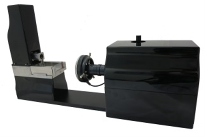
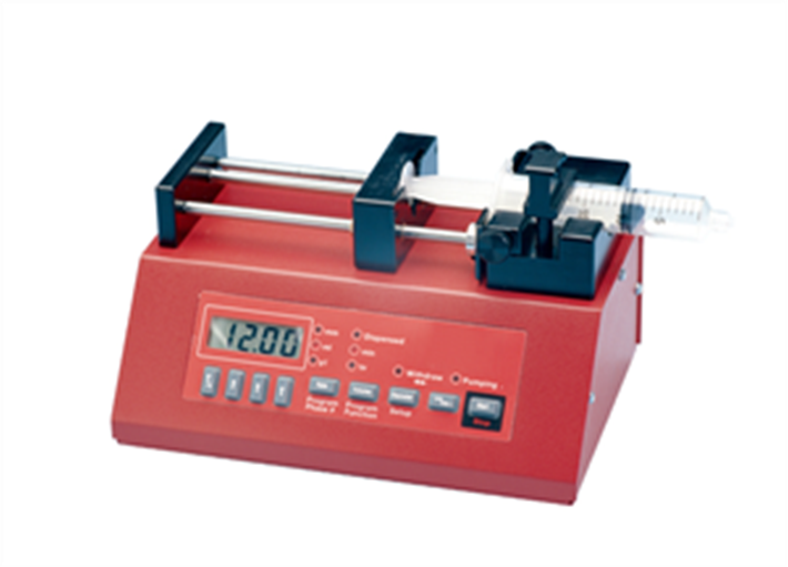
The MicroSquisher is designed to perform compression testing on specimens between 50 and 2000µm with force resolutions as small as 0.05µN.
A micro-scale tension/compression test system
The MicroSquisher does what others can’t. Smaller specimens, better force resolution, easier test setups, and great visuals. It operates on the same principles as an atomic force microscope – cantilever mechnanics – but at much larger scale. Applications are diverse but include small tissue samples, hydrogel microspheres, cell spheroids, and engineered tissue growth matrix.
The MicroSquisher’s setup, operation and data collection software module allows for simple execution of standard or customized test protocols. Test parameters such as displacement magnitudes, durations and data/image collection rates are specified in a table format for quick access and modification. A template system is used to quickly reload the desired test parameters once a protocol has been established.
While the test is running, the software provides real-time results graphing and a live video feed to facilitate user monitoring of the test progress. Ask anyone who has used our equipment and they will be quick to tell you how intuitive and useful this software package really is.
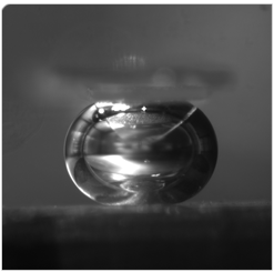
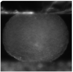
Specimens & Mounting
 |
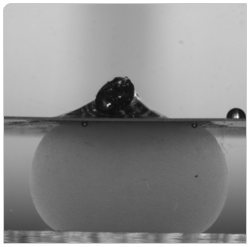 |
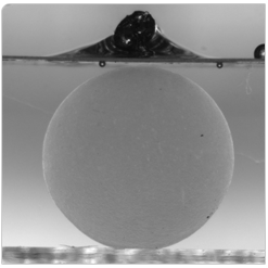
| Uncompressed and compressed hydrogel microsphere. Original diameter: 3mm Compression: 40% | |
 |
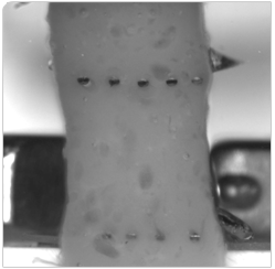 |
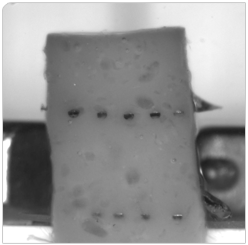
| Unstretched and stretched gel scaffold Original size: 3.3mm wide, 1.6mm thick Extension: 40% Mounting via 5 puncture pins at 0.7mm spacing Modulus was 490Pa | |
 |
|
| Compressed air bubble Approximate size: 0.6mm |
Compressed cell spheroid Approximate size: 500μm |
Videos
MicroScale Mechanical Testing Examples
Mechanical Testing of Soft Gels using the CellScale MicroSquisher
Muscle Tissue Construct Testing at Kent State University
CellScale MicroSquisher Instructional Overview
Compression Test of a Hydrogel Microsphere
Hydrogel Tension Test
Zebrafish Embryo Cell Spheroid Compression Testing
This will be the feed from an external API...
Bekesi, N., Dorronsoro, C., de la Hoz, A., & Marcos, S. (2016). Material Properties from Air Puff Corneal Deformation by Numerical Simulations on Model Corneas. PLOS ONE, 11(10), e0165669. http://doi.org/10.1371/journal.pone.0165669
Gillies, D., Gamal, W., & Downes, A. (2017). Real-time and non-invasive measurements of cell mechanical behaviour with optical coherence phase microscopy. Of SPIE Vol. http://doi.org/10.1117/12.2251492
Hached, F., Vinatier, C., Pinta, P.-G., Hulin, P., Le Visage, C., Weiss, P., … Grimandi, G. (2017). Polysaccharide Hydrogels Support the Long-Term Viability of Encapsulated Human Mesenchymal Stem Cells and Their Ability to Secrete Immunomodulatory Factors. Stem Cells International, 2017, 1–11. http://doi.org/10.1155/2017/9303598
Henry, N., Clouet, J., Fragale, A., Griveau, L., Chédeville, C., Véziers, J., … Le Visage, C. (2017). Pullulan microbeads/Si-HPMC hydrogel injectable system for the sustained delivery of GDF-5 and TGF-β1: new insight into intervertebral disc regenerative medicine. Drug Delivery, 24(1), 999–1010. http://doi.org/10.1080/10717544.2017.1340362
Kasukonis, B. M., Kim, J. T., Washington, T. A., & Wolchok, J. C. (2016). Development of an infusion bioreactor for the accelerated preparation of decellularized skeletal muscle scaffolds. Biotechnology Progress, 32(3), 745–755. http://doi.org/10.1002/btpr.2257
Kvasnytsia, M., Famaey, N., Böhm, M., & Verhoelst, E. (2016). Patient Specific Vascular Benchtop Models for Development and Validation of Medical Devices for Minimally Invasive Procedures. Journal of Medical Robotics Research, 1(3), 1640008. http://doi.org/10.1142/S2424905X16400080
Mironov, V., & Yu, H. (2017). Tensiometric estimation of material properties of tissue spheroids. Retrieved from http://dc.engconfintl.org/biofab_tissue_model/18/
Monaco, L. A., DeWitte-Orr, S. J., & Gregory, D. E. (2016). A comparison between porcine, ovine, and bovine intervertebral disc anatomy and single lamella annulus fibrosus tensile properties. Journal of Morphology, 277(2), 244–251. http://doi.org/10.1002/jmor.20492
Park, D. W., Sebastiani, A., Yap, C. H., Simon, M. A., & Kim, K. (2016). Quantification of Coupled Stiffness and Fiber Orientation Remodeling in Hypertensive Rat Right-Ventricular Myocardium Using 3D Ultrasound Speckle Tracking with Biaxial Testing. PLOS ONE, 11(10), e0165320. http://doi.org/10.1371/journal.pone.0165320
Pradhan, S., Clary, J. M., Seliktar, D., & Lipke, E. A. (2017). A three-dimensional spheroidal cancer model based on PEG-fibrinogen hydrogel microspheres. Biomaterials, 115, 141–154. http://doi.org/10.1016/J.BIOMATERIALS.2016.10.052
Pradhan, S., Hassani, I., Seeto, W. J., & Lipke, E. A. (2017). PEG-fibrinogen hydrogels for three-dimensional breast cancer cell culture. Journal of Biomedical Materials Research Part A, 105(1), 236–252. http://doi.org/10.1002/jbm.a.35899
Seeto, W. J., Tian, Y., Winter, R. L., Caldwell, F. J., Wooldridge, A. A., & Lipke, E. A. (2017). Encapsulation of Equine Endothelial Colony Forming Cells in Highly Uniform, Injectable Hydrogel Microspheres for Local Cell Delivery. Tissue Engineering Part C: Methods, 23(11), 815–825. http://doi.org/10.1089/ten.tec.2017.0233
Silva, K. R., Rezende, R. A., Pereira, F. D. A. S., Gruber, P., Stuart, M. P., Ovsianikov, A., … Mironov, V. (2016). Delivery of Human Adipose Stem Cells Spheroids into Lockyballs. PLOS ONE, 11(11), e0166073. http://doi.org/10.1371/journal.pone.0166073
Sivakumaran, D., Mueller, E., & Hoare, T. (2017). Microfluidic production of degradable thermoresponsive poly( N -isopropylacrylamide)-based microgels. Soft Matter, 13(47), 9060–9070. http://doi.org/10.1039/C7SM01361B
Stewart, D. M., Monaco, L. A., & Gregory, D. E. (2017). The aging disc: using an ovine model to examine age-related differences in the biomechanical properties of the intralamellar matrix of single lamellae. European Spine Journal, 26(1), 259–266. http://doi.org/10.1007/s00586-016-4603-4
Wu, S., Wang, Y., Streubel, P., & Duan, B. (2017). Living nanofiber yarn-based woven biotextiles for tendon tissue engineering using cell tri-culture and mechanical stimulation. Acta Biomaterialia. Retrieved from http://www.sciencedirect.com/science/article/pii/S1742706117305573
Yu, C., Kornmuller, A., Brown, C., Hoare, T., & Flynn, L. E. (2017). Decellularized adipose tissue microcarriers as a dynamic culture platform for human adipose-derived stem/stromal cell expansion. Biomaterials, 120, 66–80. http://doi.org/10.1016/J.BIOMATERIALS.2016.12.017


Request
Catalogue
Chat
Print