




Celloger Mini Plus
Automated Live Cell Imaging system with bright field and options for green or red fluorescence
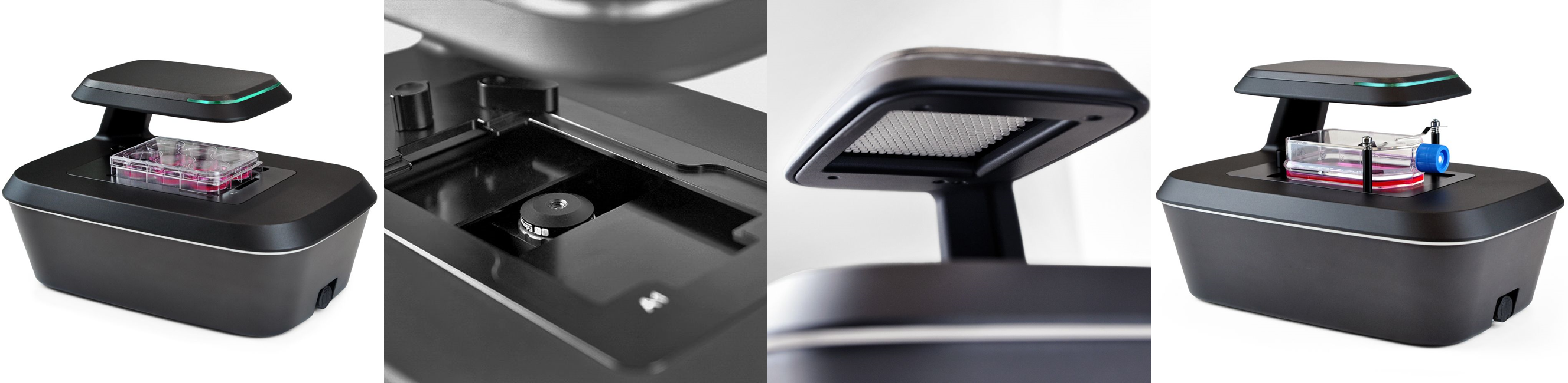
|
|
 Celloger Min Plus Instruction Manual
Automated Live Cell Imaging System
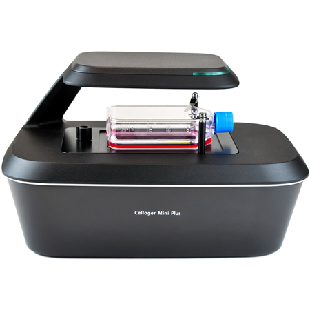
Celloger Mini Plus is an automated live cell imaging system that is equipped with an advanced fluorescence and bright field microscopy, autofocusing and real time multi-position imaging technology for a well plate, dish or T-flask.
The streamlined process provides an easy workflow solution giving you a full set of tools you need to acquire the best quality images and accurate research results.
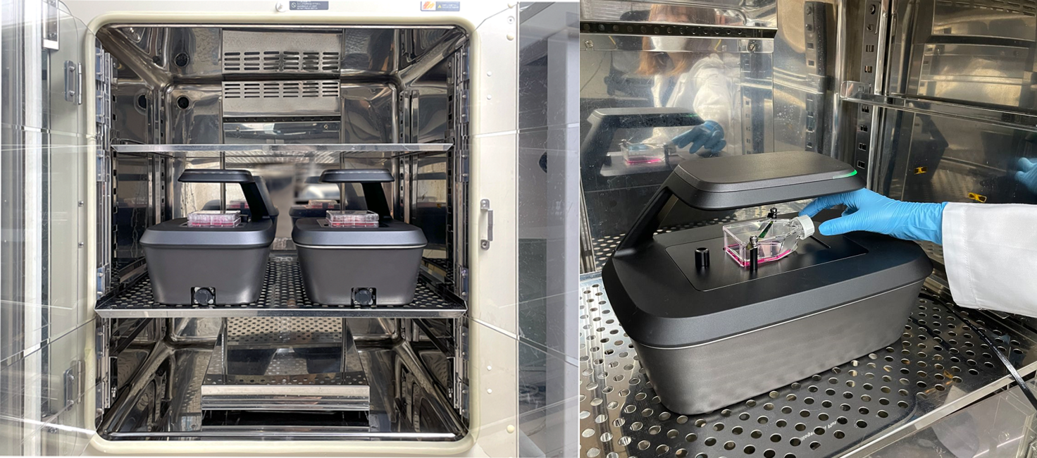
The compact nature of the Celloger Mini Plus allows positioning in an incubator providing improved cell viability as there is less disturbances over the course of your experiment reducing chances of cellular abnormality.
The Celloger Mini Plus live cell imaging system includes two software packages
- Celloger Mini Plus Scan to control the actual operation of Celloger Mini Plus device which includes real-time cell monitoring and time-lapse imaging functions
- Celloger Mini PlusAnalysis for analyzing and post-processing the images. This comes with an unlimited software usage license, ideal for allowing multiple users to work with the unit without additional fees
Expand Your Cell Discoveries with Automated Live Cell Imaging
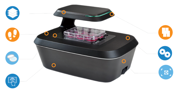
 |
Compact size |
 |
Multi-position imaging |
 |
Autofocusing |
 |
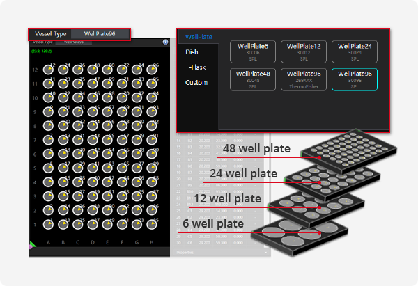
High compatibility |
 |
Intuitive interface |
 |
Z-stacking imaging |
 |
Image stitching |
Unique Benefits of Celloger Mini Plus
Stable Stage – Get clearer images with a stable plate. Unlike other devices, Celloger Mini Plus has a fixed stage and the optics move.
Open-Source Data – Unlike other brands, Celloger Mini Plus gives you the raw images so that you can work with any analysis software you prefer, like Image J.
Regular Software Updates – Stay current with all the new functions of the Celloger Mini Plus software. Customers are lifetime members and are never charged for the software updates.
Unlimited Software Usage License – Multiple users may analyze images using their own computers (after the scanning is complete) without additional fees. Other brands limit usage to one computer.
Specifications
| Type | Description |
|---|---|
| Dimension | 226 x 358 x 215 mm |
| Weight | 5.6kg |
| Objective Lens | 2X or 4X or 10X depending on model (see options) |
| Imaging modes | Bright field with optional Green Fluorescence or Red Fluorescence depending on model (see options) |
| Light source | LED |
| Camera | 5MP CMOS |
| Optics Stage | Motorized XYZ |
| Imaging positions | Multiple |
| File export format | TIFF, AVI, JPEG, PNG |
| Culture vessels | Well plate (6, 12, 24, 48, 96 wells), Dish* (35 mm, 60 mm, 90 mm), and T-flask* (25 cm2 , 75cm2) |
| Operating environment | 10~40°C, 20~95% humidity |
| Power requirements | 100-240V, ~50/60Hz |
| Output ports | Ethernet |
| Software** | Celloger Mini Plus Scan and Celloger Mini Plus Analysis |
| Accessories | PoE adapter, ethernet cable, USB/LAN adapter, USB memory |
| Warranty | 1 year |
* Requires optional adapters
** Requires External PC/Laptop with Windows 11 (not included with system). Recommended specs for PC/Laptop: Storage >1TB; Monitor: 1920 x 1080 pixels (Full HD)
Options
| Order code | Description |
|---|---|
| EVI-LCI-01-18 | Celloger Mini Plus, Brightfield, 4X Magnification |
| EVI-LCI-01-19 | Celloger Mini Plus, Brightfield, 10X Magnification |
| EVI-LCI-01-20 | Celloger Mini Plus, Brightfield with Green Fluorescence, 4X Magnification |
| EVI-LCI-01-21 | Celloger Mini Plus, Brightfield with Green Fluorescence, 10X Magnification |
| EVI-LCI-01-22 | Celloger Mini Plus, Brightfield with Red Fluorescence, 4X Magnification |
| EVI-LCI-01-23 | Celloger Mini Plus, Brightfield with Red Fluorescence, 10X Magnification |
Features
Multipoint imagingThe optics system travels 117mm x 77mm, x and y axis respectively, multiple points within the travel range can be captured following the schedule (intervals, cycles, total time) set by the researcher. Field of view: - 4X: 1.345 x 1.017mm - 10X: 0.574 x 0.43mm |
 |
|
|
Stable imaging performanceCelloger Mini Plus doesn’t have a moveable stage but instead, the camera located inside the system moves to capture the images of cell in multiple positions |
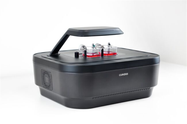
Compact sizeCelloger Mini Plus is compact in size with 226(h) x 358(l) x 215(w) mm where several Celloger Mini Plus systems can fit into a standard CO2 incubator. |
 |
Scanning Application
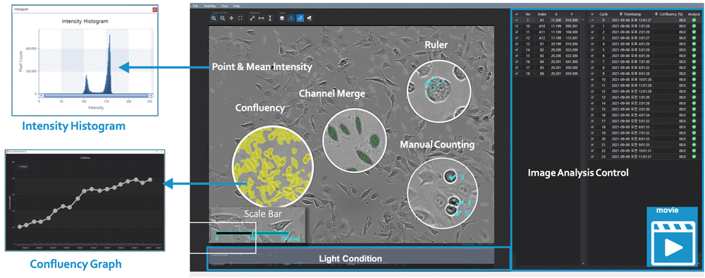
The scanning application is used for capturing images. You can preview cells, schedule image capture, adjust light and contrast, and monitor time lapse progression from one intuitive screen. It includes auto-focusing technology that finds a clear focal plane of cells and has excellent repeatability.
Z-Stacking
With the Z-stacking function, where images of multiple planes of focus are merged, spheroid cells can be clearly observed under time-lapse imaging.
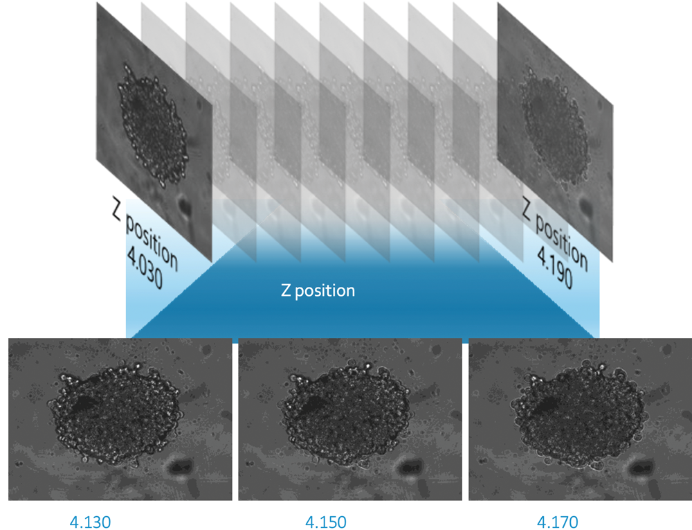
Image Stitching
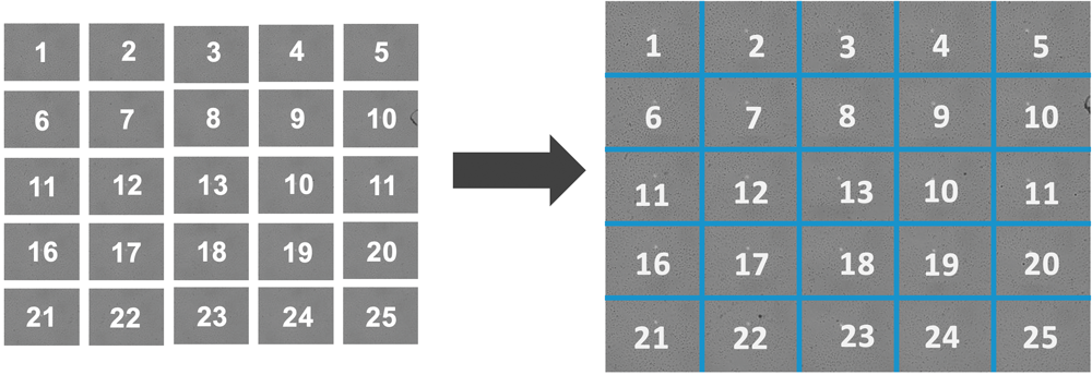
Image stitching lets you capturing multiple images and combine the overlapping parts to enable high-resolution mapping of a large sample area.
Time Lapse Imaging
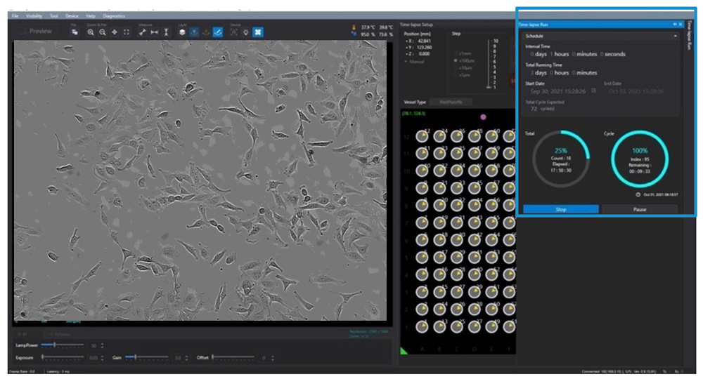
The Celloger Mini Plus Scan app gives you an intuitive interface for scheduling your time-lapse images. You can set the total time, cycle and intervals for the time lapse images of your live cell imaging setup.
Analysis Application
Celloger Mini Plus Analysis has a variety of tools in the analysis application simplify the analytic process, reducing errors and saving time.
- Intensity Histograms
- Confluency Graphs
- Channel Merge
- Video Creation
- Time Lapse Imaging
- Manual Counting
- Measurement
- Z-Stacking Image
- More
Celloger Mini Plus - DEMO
Request a demo of the Celloger Mini Plus live cell imaging system by adding to the basket and checking out.
One of our technical specialists will contact you to arrange a demonstration.
EVI-LCI-01-18
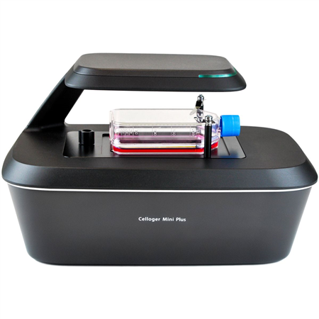
Celloger Mini Plus, Brightfield, 4X Magnification Automated Live Cell Imaging System
- Objective Lens: 4X
- Imaging Modes: Bright Field
- Output: Ethernet via PoE Adapter (included)
- Software: Celloger Mini Plus Scan and Celloger Mini Plus Analysis (Requires Windows 11 PC/Laptop - not included)
EVI-LCI-01-19
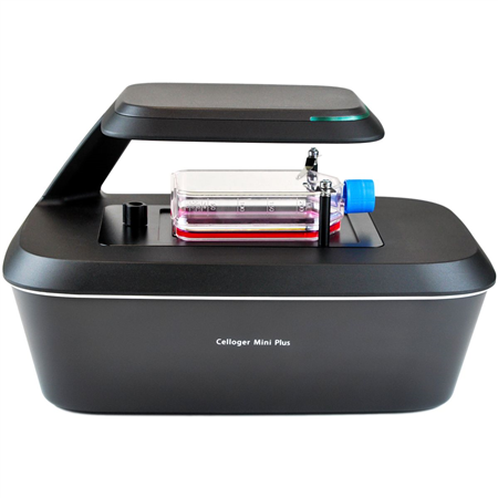
Celloger Mini Plus, Brightfield, 10X Magnification Automated Live Cell Imaging System
- Objective Lens: 10X
- Imaging Modes: Bright Field
- Output: Ethernet via PoE Adapter (included)
- Software: Celloger Mini Plus Scan and Celloger Mini Plus Analysis (Requires Windows 11 PC/Laptop - not included)
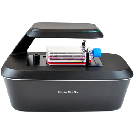
EVI-LCI-01-20
Celloger Mini Plus, Brightfield & Green Fluorescence, 4X Magnification Automated Live Cell Imaging System
- Objective Lens: 4X
- Imaging Modes: Bright Field, Green Fluorescence
- Output: Ethernet via PoE Adapter (included)
- Software: Celloger Mini Plus Scan and Celloger Mini Plus Analysis (Requires Windows 11 PC/Laptop - not included)
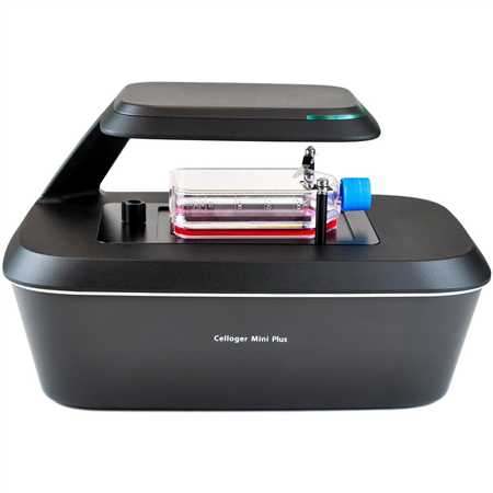
EVI-LCI-01-21
Celloger Mini Plus, Brightfield & Green Fluorescence, 10X Magnification Automated Live Cell Imaging System
- Objective Lens: 10X
- Imaging Modes: Bright Field, Green Fluorescence
- Output: Ethernet via PoE Adapter (included)
- Software: Celloger Mini Plus Scan and Celloger Mini Plus Analysis (Requires Windows 11 PC/Laptop - not included)
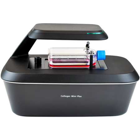
EVI-LCI-01-22
Celloger Mini Plus, Brightfield & Red Fluorescence, 4X Magnification Automated Live Cell Imaging System
- Objective Lens: 4X
- Imaging Modes: Bright Field, Red Fluorescence
- Output: Ethernet via PoE Adapter (included)
- Software: Celloger Mini Plus Scan and Celloger Mini Plus Analysis (Requires Windows 11 PC/Laptop - not included)
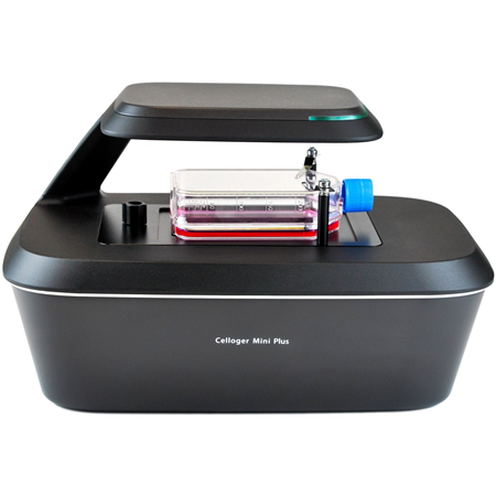
EVI-LCI-01-23
Celloger Mini Plus, Brightfield & Red Fluorescence, 10X Magnification Automated Live Cell Imaging System
- Objective Lens: 10X
- Imaging Modes: Bright Field, Red Fluorescence
- Output: Ethernet via PoE Adapter (included)
- Software: Celloger Mini Plus Scan and Celloger Mini Plus Analysis (Requires Windows 11 PC/Laptop - not included)
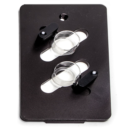
505628
Celloger Mini Plus Adapter for 35 mm FluoroDish, Dual
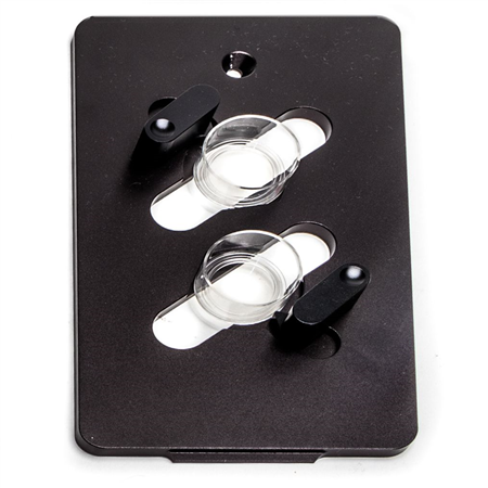
505629
Celloger Mini Plus Adapter for 60 mm Dish, Dual
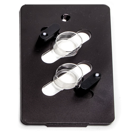
505630
Celloger Mini Plus Adapter for 90 mm Dish
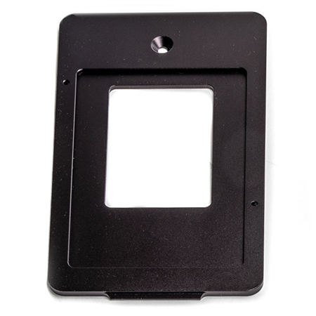
505626
Celloger Mini Plus Adapter for T-Flask 25 cm2, Single
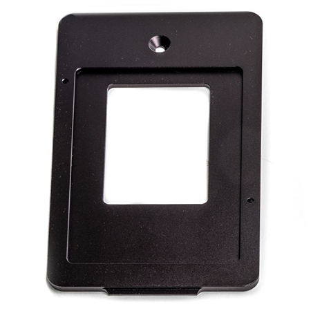
505627
Celloger Mini Plus Adapter for T-Flask 75 cm2, Single
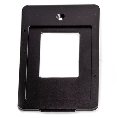
505632
Celloger Mini Plus Adapter for T-Flask 25 cm2, Dual


Request
Catalogue
Chat
Print